
Introduction
Continual myeloid leukemia (CML) is a hematological neoplasm initiated by the fusion gene BCR-ABL (1). The introduction of tyrosine kinase inhibitors (TKIs), comparable to imatinib, has considerably enhanced therapeutic efficacy for CML sufferers whereas considerably bettering their prognosis (2). Nonetheless, intricate escape mechanisms employed by tumor cells inevitably hinder the effectiveness of those kinase medicine and result in the gradual growth of drug resistance in sufferers with CML (3). These resistance mechanisms embody each main and secondary elements; amongst them, mutations in BCR-ABL protein play a vital position (4). Regardless of the developments achieved within the growth of novel TKIs that focus on particular mutation websites related to enhanced remedy response in CML (5); challenges persist as a result of rising new mutation websites over time in addition to non-mutation-based resistance mechanisms that come up throughout remedy course (6). Due to this fact, a extra complete evaluation of the molecular biology and metabolic traits of CML cells holds vital scientific worth for remedy decision-making and prognosis analysis in sufferers with CML.
Ferroptosis is a novel type of cell loss of life, characterised by distinct mechanisms and morphology in comparison with apoptosis, necrosis, and autophagy (7). The method is initiated by intracellular divalent iron or ester oxygenase, ensuing within the peroxidation of extremely expressed unsaturated fatty acids on the cell membrane and subsequent induction of ferroptosis (8–10). Morphological adjustments noticed in cells present process ferroptosis embody disruption of the cell membrane, mitochondrial outer membrane, and lack of cristae (11). The prevalence of ferroptosis entails varied regulatory pathways such because the classical GPX4-regulated mechanism (Cyst(e)ine/GSH/GPX4 axis) (12), in addition to GPX4-independent mechanisms like NAD(P)H/FSP1/CoQ10 axis (13), GCH1/BH4/DHFR axis (14), and squalene accumulation. Moreover, signaling pathways together with E-cadherin-NF2-Hippo-YAP, AMPK, and HIF2α-HILPDA additionally modulate mobile sensitivity to ferroptosis (15–17). Quite a few research have demonstrated that focused induction of ferroptosis holds promise as a brand new therapeutic technique for acute myeloid leukemia (18–20). Liu et al.’s analysis revealed TXNRD1’s essential position in cysteine depletion-induced ferroptosis in CML cells in vitro (18, 21). Nonetheless, there stays a restricted understanding relating to the connection between ferroptosis and CML, as properly as its underlying mechanism, necessitating additional complete investigation.
On this research, we performed a complete evaluation of the ferroptosis pathway and gene expression traits in CML, aiming to elucidate the underlying mechanism of ferroptosis and its interplay with the CML tumor microenvironment. By means of multi-group cohort evaluation, we validated the diagnostic worth of ferroptosis-related genes (FRGs) in CML, and subsequent experiments additional confirmed the potential therapeutic significance of concentrating on ferroptosis in overcoming drug resistance.
Strategies
Information acquisition and preprocessing
The sequencing information of CML cohorts GSE13159, GSE144119, GSE4170, and GSE44589 had been obtained from the Gene Expression Omnibus (GEO) database. The evaluation cohort for this mission was the GSE13159 cohort, which consisted of 76 CML samples and 74 regular samples. Uncooked sequencing information had been downloaded and normalized for subsequent evaluation. The validation cohort (GSE144119) included 48 newly recognized CML samples, 32 remission CML samples, and 17 regular samples that had been transformed to transcripts per kilobase million (TPM) values. For scientific validation functions, transcriptome sequencing was carried out on 5 chronic-phase CML samples, 5 blast disaster samples, and 5 regular management samples with written consent from sufferers accepted by the Ethics Committee of the Second Affiliated Hospital of Nanchang College; these information had been additionally reworked into TPM values for additional validation. To distinguish between different forms of leukemia comparable to acute lymphoblastic leukemia (750 instances), acute myeloid leukemia (542 instances), persistent lymphocytic leukemia (448 instances), and myelodysplastic syndromes (206 instances), a subset of the GSE13159 cohort was utilized. Moreover, we used the imatinib-treated pattern dataset from GSE44589 containing 198 sequenced samples to guage remedy response in CML sufferers. Moreover, single-cell RNA-seq information from the GES76312 cohort had been employed to visualise clusters utilizing the uniform manifold approximation and projection (UMAP) algorithm. Lastly, we retrieved ferroptosis pathway genes from the MSigDB database (https://www.gsea-msigdb.org/gsea/msigdb/index.jsp).
Differential expression evaluation of FRG
The “limma” software program package deal was employed for conducting differential expression evaluation of FRG. Adjusted p values under 0.05 had been thought-about vital, indicating the presence of differentially expressed FRGs (DEFRGs) between CML and regular samples. Subsequently, we carried out Gene Ontology (GO) annotation and Kyoto Encyclopedia of Genes and Genomes (KEGG) pathway enrichment evaluation on these genes utilizing the “clusterProfiler” package deal (22). To quantify the exercise of a organic pathway or gene set, we utilized the Gene Set Variation Evaluation (GSVA) algorithm to calculate an enrichment rating (23).
Correlation evaluation and protein-protein interplay (PPI) community development
The Spearman technique was employed for correlation evaluation. The STRING database (https://string-db.org/) was used to investigate the PPI of DEFRG. Subsequently, the PPI community was visualized utilizing Cytoscape software program.
Evaluation of immune cell infiltration
The estimation of immune cell infiltration was performed by using the deconvolution algorithm “CIBERSORT” to precisely quantify the proportions of twenty-two distinct immune cell varieties based mostly on the gene expression profiles of particular person samples (24).
Potential regulatory mechanisms related to ferroptosis
Weighted correlation community evaluation (WGCNA) was employed to establish the genes related to ferroptosis scores within the GSE13159 cohort (25). Pearson correlation evaluation was utilized to assemble the adjacency matrix for all matched genes, and the scale-free topology of this matrix was established based mostly on an optimum mushy threshold energy. Subsequently, the adjacency matrix was reworked right into a topological overlap matrix (TOM). By using the TOM dissimilarity measure, modules consisting of genes exhibiting related expression patterns had been recognized by way of common linkage hierarchical clustering, with a minimal module dimension set at 30 and a reduce top at 0.2. Lastly, an analysis of the correlation between module signature genes (MEs) and ferroptosis rating was carried out.
Evaluation of the diagnostic worth of FRGs
To establish diagnostic biomarkers for CML, three machine studying algorithms, particularly help vector machine recursive function elimination (SVM-RFE), least absolute shrinkage choice operator (LASSO), and random forest (RF) had been employed to display the diagnostic FRGs. Moreover, LASSO regression evaluation was used to calculate regression coefficients for the diagnostic FRGs, and a CML danger rating diagnostic mannequin was constructed utilizing the next system:
the place i represents the particular diagnostic FRG and “Coef” and “ExpGene” denote the regression coefficient and expression worth of that specific FRG respectively. By setting up this danger rating mannequin, we will additional assess the mixed diagnostic worth of FRGs.
Revealing molecular subtypes by way of FRG expression profiling
To comprehensively assess inter-individual variations in CML sufferers, we employed the “ConsensusClusterplus” package deal to conduct a cluster evaluation of CML samples based mostly on the expression profiles of the diagnostic FRGs, aiming to establish distinct molecular subtypes inside CML (26). The robustness and stability of the clustering outcomes had been confirmed by way of 1000 iterations. Moreover, principal part evaluation (PCA) was utilized for classification validation.
Prediction of the sensitivity of CML samples to TKI remedy and immunotherapy
The expression matrix and drug response information of blood cell strains from the Most cancers Genome Mission (CGP) database had been utilized on this research to foretell the half-maximal inhibitory concentrations (IC50) of CML samples to TKIs. This prediction was made utilizing the “pRRophetic” package deal, a computational device generally used for such analyses (27). To additional examine the response of various danger rating teams in the direction of anti-PD-1 and anti-CTLA4 immune checkpoint inhibitors, we employed the “SubMap” algorithm out there at a publicly accessible web site referred to as GenePattern. The SubMap algorithm is well known for its capability to forecast remedy responses based mostly on gene expression profiles. To evaluate the extent of immune escape exhibited by tumor cells in CML samples, we computed the TIDE rating utilizing a longtime on-line useful resource often called Tumor Immune Dysfunction and Exclusion (TIDE).
Development of microRNA (miRNA) regulatory community for diagnostic FRGs
We employed miRTarBase, miRDB, and TargetScan databases to foretell the binding websites of miRNAs on CML diagnostic ARGs. Subsequently, we filtered out the miRNA-target pairs that had been predicted by all three databases. The GSE90773 cohort was utilized to establish differentially expressed miRNAs between CML cells and regular cells, which served as the idea for setting up the miRNA regulatory community.
In vitro experiments
The CML cell line K562 was cultured in RPMI1640 medium supplemented with 10% fetal bovine serum and 1% penicillin-streptomycin in a humidified incubator saturated with 5% CO2 at 37°C. The K562 cells had been uncovered to imatinib, and the focus was progressively elevated till the event of K562/IR cells able to sustained development in a medium containing 1μM of imatinib. This focus is taken into account physiologically related and will simulate the height plasma/serum degree of imatinib (5μM). Transcriptome sequencing evaluation was performed on K562, K562/IR, K562/IR management, and erastin-treated K562/IR cells. The processing process employed on this research was based mostly on our earlier analysis (28). The focus of imatinib was progressively elevated till the induction of resistant cells was accomplished. Cell viability was assessed utilizing the cell counting kit-8 (CCK-8) assay. For this assay, 5-e3 cells had been seeded in 96-well plates, and every group was repeated thrice. After the indicated tradition time, 10 μL of CCK8 resolution was added, adopted by incubation at 37°C for two hours. The optical density (OD) worth at 450 nm was measured utilizing a microplate reader. Apoptosis detection concerned staining cells with the Annexin V-PE/7-AAD apoptosis detection package and subsequent examination in a stream cytometer. Moreover, reactive oxygen species (ROS) had been detected utilizing a fluorescent probe DCFH-DA in stream cytometry. The degrees of GSH and GSSH had been decided utilizing Solarbio’s BC1175 and BC1185 kits, respectively. Bioss’ AK091 package was used for GPX4 exercise measurement. All reagents had been employed following the producer’s directions. Cell homogenization was carried out utilizing lysate buffer to facilitate the response between REDOX substances within the pattern and reagents, ensuing within the formation of adducts that may be quantified by way of colorimetry.
Statistical evaluation
All analyses had been performed utilizing the R software program and corresponding software program packages. Variations between two or extra teams had been assessed utilizing the Wilcoxon rank sum take a look at and the Kruskal-Wallis take a look at, respectively. The diagnostic worth of biomarkers was decided by way of receiver working attribute (ROC) curve evaluation. A bilateral P-value lower than 0.05 signifies a statistically vital distinction.
Outcomes
Molecular traits linked to ferroptosis in CML
We performed a complete analysis of ferroptosis exercise and molecular traits in CML utilizing transcriptomics evaluation. The GSVA algorithm was utilized to calculate ferroptosis scores, revealing considerably decrease ferroptosis scores in CML samples in comparison with regular samples (Determine 1A), whereas the ferroptosis rating elevated following remedy remission (Determine 1B). Sufferers in blast disaster (BC) exhibited even decrease ferroptosis scores than these within the persistent section (CP) (Figures 1C, D) (As a result of restricted pattern dimension and vulnerability to particular person outliers, though Determine 1C doesn’t exhibit a statistically vital distinction, the general pattern persists that BC sufferers show decrease ferroptosis scores in comparison with CP sufferers.), and people with main molecular responses displayed increased ferroptosis scores in comparison with non-responders (Determine 1E). Single-cell evaluation constantly demonstrated a pattern of decreased ferroptosis scores in CML sufferers, notably these in BC, which subsequently elevated after remedy with TKI (Figures 1F-H). Differential expression evaluation indicated the down-regulation of quite a few genes related to ferroptosis in CML samples (Figures 1I, J), together with these concerned in iron ion homeostasis, mitochondrial outer membrane perform, and ligase exercise (Determine 1K). These differentially expressed genes had been primarily enriched in signaling pathways associated to ferroptosis, metabolic pathways, mineral absorption, and cysteine and methionine metabolism (Determine 1L). PPI community evaluation recognized STEAP3, TFRC NCQA4 TP53 IREB2 as hub genes inside the community fashioned by these DEFRG (Determine 1M). Volcano plot evaluation additional revealed down-regulation of gene expression for varied suppressors of ferroptosis in CML samples (Figures 1N, O). Due to this fact, we speculate that the noticed decrease ferroptosis rating in CML could also be attributed to an general lower in inhibition of this course of inside most cancers cells indicating their heightened susceptibility in the direction of present process cell loss of life by way of the mechanism of the ferroptosis pathway. Ferroptosis is intently linked to lipid metabolism, and our findings reveal a big improve within the exercise of unsaturated fatty acids comparable to linoleic acid, arachidonic acid, and α-linolenic acid in CML (Determine 1P). Contemplating that the peroxidation of unsaturated fatty acids is a prerequisite for ferroptosis to happen, this end result additional helps the speculation that CML reveals heightened susceptibility to ferroptosis. These outcomes collectively point out an aberrant regulation of ferroptosis in CML samples, which can have implications for the initiation and development of the illness.
Determine 1 The traits of ferroptosis rating and FRG expression in CML samples. (A-E) Variations in ferroptosis scores between CML samples and regular samples had been noticed in varied datasets: (A) GSE13159, (B) GSE144119, (C) our scientific cohort, (D) GSE4170, (E) GSE44589. (F-H) UMAP evaluation of the CML single-cell sequencing dataset GSE76312 revealed the distribution of ferroptosis scores amongst completely different sufferers. (I-J) Volcano map (I) and warmth map (J) illustrated the expression traits of FRG. (Okay, L) Useful annotation (Okay) and pathway enrichment evaluation (L) had been performed on DEFRG. (M) PPI community evaluation was carried out on DEFRG. (N, O) Expression traits of ferroptosis suppressors and drivers had been examined. (P) Variations in lipid metabolic pathway scores between regular and CML samples. BC refers to blast disaster, CP to persistent section, MMR to main molecular response, and NR to no response. *p < 0.05; **p < 0.01; ***p < 0.001.
The correlation between the ferroptosis rating and the immune microenvironment in addition to signaling pathways
The connection between the ferroptosis rating and the immune microenvironment of CML in addition to most cancers pathways was additional analyzed. It was noticed that there have been vital associations between the ferroptosis rating and key tumor marker pathways, together with xenobiotic metabolism, reactive oxygen species pathway, heme metabolism, and epithelial mesenchymal transition actions (Determine 2A). Furthermore, optimistic correlations had been discovered with glycolysis and hypoxia, whereas detrimental correlations had been noticed with Notch signaling and WNT beta-catenin signaling. These findings counsel that an elevated exercise within the ferroptosis pathway is accompanied by enhanced most cancers cell metabolism. Immune infiltration evaluation revealed a optimistic correlation between the ferroptosis rating and eosinophil infiltration, M0 macrophage infiltration, in addition to regulatory T cell (Treg) infiltration; in the meantime, a detrimental correlation was recognized with naive CD4+ T cells (Determine 2B). Moreover, a big optimistic correlation was additionally discovered between the ferroptosis rating and gene expression of immune checkpoints LAG3 and TNFRSF9 (Determine 2C), indicating potential immunosuppression amongst sufferers with excessive ferroptosis scores.
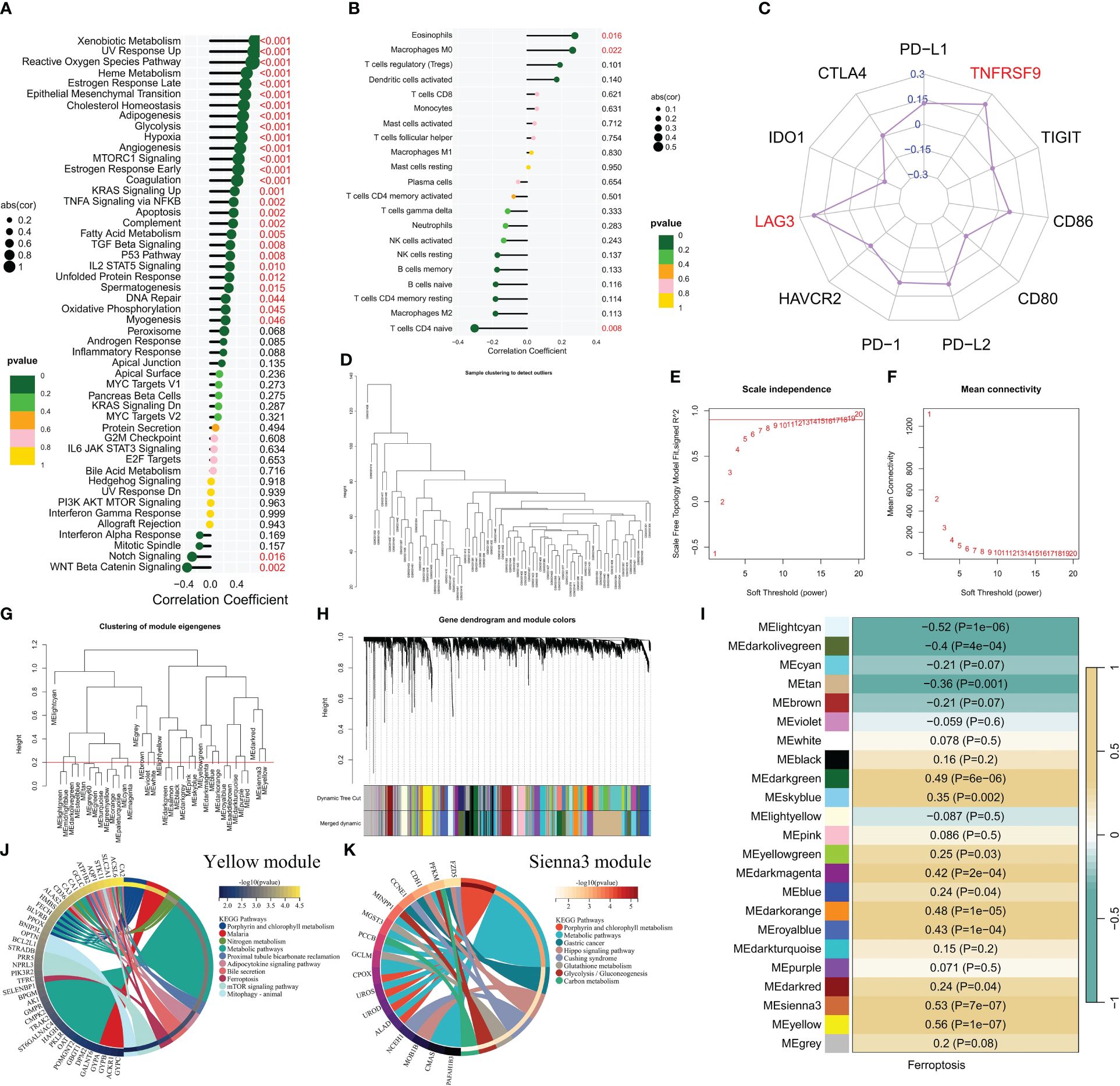
Determine 2 The correlation between the ferroptosis rating and the immune microenvironment and signaling pathways. (A-C) Correlation evaluation revealed associations between the ferroptosis rating and enrichment scores of tumor marker gene units (A), infiltration of immune cells (B), and expression of immune checkpoints (C). (D) Cluster plot displaying CML samples. (E, F) Scale-free becoming index and common connectivity had been used to investigate varied mushy threshold powers. (G) Clustering was carried out on completely different modules, with a reducing top set at 0.2 represented by the crimson line. (H) Cluster plots had been generated based mostly on completely different measures utilizing 1-TOM calculation. (I) Heatmap illustrating the correlation between module genes and ferroptosis rating. (J, Okay) KEGG enrichment evaluation was performed for yellow module genes, in addition to sienna3 module genes.
To achieve a deeper understanding of the underlying mechanisms related to ferroptosis in CML, we performed WGCNA to discover the community of co-expressed genes considerably correlated with ferroptosis scores. The cluster dendrogram depicted the clustering traits of all CML samples (Determine 2D). Figures 2E, F illustrate the scale-free match exponential and common connectivity evaluation for varied mushy threshold powers. We set the reduce top at 0.2 to incorporate modules exhibiting a correlation coefficient larger than 0.8 (Determine 2G). Primarily based on an optimum mushy threshold energy β=15 (unscaled R^2 = 0.9), WGCNA categorised the highest 5000 genes with the best normal deviation into 23 impartial co-expression modules (Determine 2H). The correlograms depicting module-trait relationships revealed that each yellow and sienna3 modules exhibited robust correlations with ferroptosis scores (Determine 2I). KEGG enrichment evaluation demonstrated that these two modules had been enriched in porphyrin and chlorophyll metabolism in addition to metabolic pathways (Figures 2J, Okay). Moreover, yellow module genes had been discovered to be related to nitrogen metabolism, adipocytokine signaling pathway, mTOR signaling pathway, and mitophagy; whereas sienna3 module genes confirmed enrichment in hippo signaling pathway, glutathione metabolism, glycolysis/gluconeogenesis, and carbon metabolism. The findings counsel that metabolic reprogramming might contribute to the malignant proliferation of CML cells, whereas additionally enhancing the susceptibility of CML cells to ferroptosis by producing increased ranges of ROS and unsaturated fatty acids (11, 29).
Evaluation of the diagnostic worth of FRG
We performed additional evaluation on the diagnostic worth of FRG in CML. Three machine studying algorithms, particularly LASSO, RF, and SVM-RFE, had been employed for dimensionality discount to pick out probably the most informative FRGs. From the DEFRGs, we recognized 5, 6, and 6 variables that precisely distinguished CML samples from regular samples, respectively (Figures 3A–E). Amongst these variables, there have been 5 overlapping diagnostic FRGs (ACSL6, SLC11A2, HMOX1, SLC38A1, and AKR1C3) included amongst them (Determine 3F). The expression ranges of all 5 FRGs had been considerably downregulated in CML samples in comparison with regular samples (Determine 3G). Utilizing LASSO regression evaluation, we developed a danger rating mannequin to evaluate the mixed diagnostic worth of FRG (Determine 3H, Supplementary Desk S1). The danger rating ranges had been considerably elevated within the CML samples (Determine 3I). ROC curve evaluation revealed excessive diagnostic AUC values for ACSL6 (0.818), SLC11A2 (0.864), HMOX1 (0.782), SLC38A1(0.783), AKR1C3(0.791), in addition to for the danger rating (0.920) (Determine 3J). The mixture of those 5 FRGs additional improved their diagnostic worth.
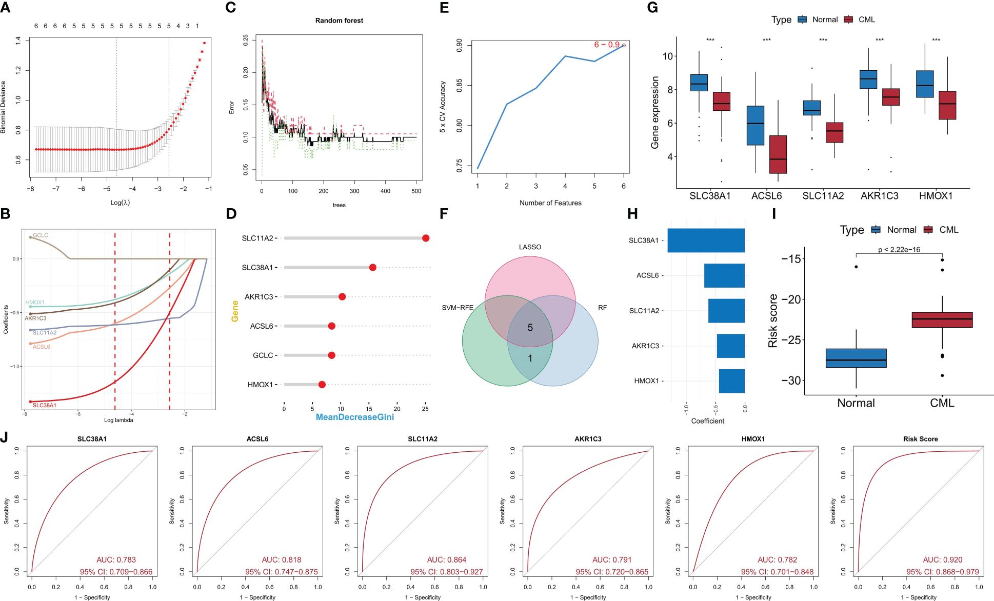
Determine 3 Identification of diagnostic FRG. (A, B) Diagnostic FRGs had been recognized by the LASSO regression algorithm. (C, D) Diagnostic FRGs had been recognized by the RF algorithm. (E) Diagnostic FRGs had been recognized by the SVM-RFE algorithm. (F) Venn diagram of variables recognized by LASSO, RF, and SVM-RFE algorithms. (G) Variations in expression of the three diagnostic FRGs between CML samples and regular samples within the GSE13159 cohort. (H) Coefficients of danger rating mannequin. (I) Variations in danger rating between CML samples and regular samples within the GSE13159 cohort. (J) ROC curve evaluation was used to guage the diagnostic worth of the 5 FRGs and danger rating within the GSE13159 cohort.
Validation of the diagnostic worth of FRG and evaluation of their position within the analysis of therapeutic impact
We confirmed the diagnostic worth of the 5 FRGS. Within the GSE144119 cohort, we noticed a big lower in expression ranges of all 5 FRGS in CML samples, which confirmed partial restoration after remedy response (Determine 4A). Moreover, the danger rating ranges had been considerably elevated in CML samples and exhibited a big lower after remedy remission (Determine 4B), thereby demonstrating the therapeutic analysis worth of FRG. ROC curve evaluation revealed that ACSL6, SLC11A2, HMOX1, SLC38A1, AKR1C3, and the danger rating mannequin had AUC values of 0.949, 0.934, 0.868, 0.842, and 0.975 respectively (Figures 4C-H); thus confirming their diagnostic worth in CML instances. In our clinically impartial cohort research, we additionally noticed a big lower in ACSL6, SLC11A2, HMOX1, and SLC38A1 expression in CML samples whereas AKR1C3 didn’t present a big distinction as a result of small pattern dimension points (Determine 4I). The danger rating ranges had been additionally considerably elevated in CML samples (Determine 4J). ROC curve evaluation demonstrated an AUC worth of 1 for the danger rating mannequin (Determine 4K). Medical sample-based sequencing information additional verified the excessive diagnostic worth related to these 5 FRGs in CML. In conclusion, now we have recognized extremely dependable FRGs which might probably function a novel adjunctive device for scientific prognosis and remedy decision-making in sufferers with CML.

Determine 4 Validation of the diagnostic worth of the diagnostic FRG. (A, B) Variations in expression of the 5 diagnostic FRGs and danger rating between CML samples and regular samples within the GSE144119 cohort (The Kruskal-Wallis take a look at was employed for the comparability among the many three teams). (C-H) ROC curve evaluation was used to guage the diagnostic worth of the 5 FRGs and danger rating within the GSE144119 cohort. (I, J) Variations in expression of the 5 diagnostic FRGs and danger rating between CML samples and regular samples in our scientific cohort. (Okay) ROC curve evaluation was used to guage the diagnostic worth of danger rating in our scientific cohort. **p < 0.01; ***p < 0.001.
Evaluation of the differential diagnostic worth of FRG
We performed a complete evaluation to guage the differential diagnostic worth of the 5 FRGs. The GSE13159 cohort included sequencing information from 750 ALL samples, 542 AML samples, 448 CLL samples, and 206 MDS samples. Curiously, the expression ranges of most FRGs, together with SLC38A1, SLC11A2, and HMOX1, had been discovered to be decrease in CML samples in comparison with different forms of hematologic tumors. Conversely, ACSL6 exhibited increased expression ranges (Determine 5A). Moreover, subsequent calculations revealed that CML samples displayed the best danger rating (Determine 5B). ROC curve evaluation demonstrated that the danger rating successfully distinguished CML from different hematological malignancies with excessive accuracy (AUC=0.844) (Determine 5C). The diagnostic worth of FRG has been systematically evaluated, and now we have additionally endeavored to research the regulatory mechanisms governing FRG expression. On this research, our focus lies on miRNA, as we goal to assemble a miRNA regulatory community to establish potential miRNAs that would inhibit FRG expression by binding to FRG in CML cells (Determine 5D).
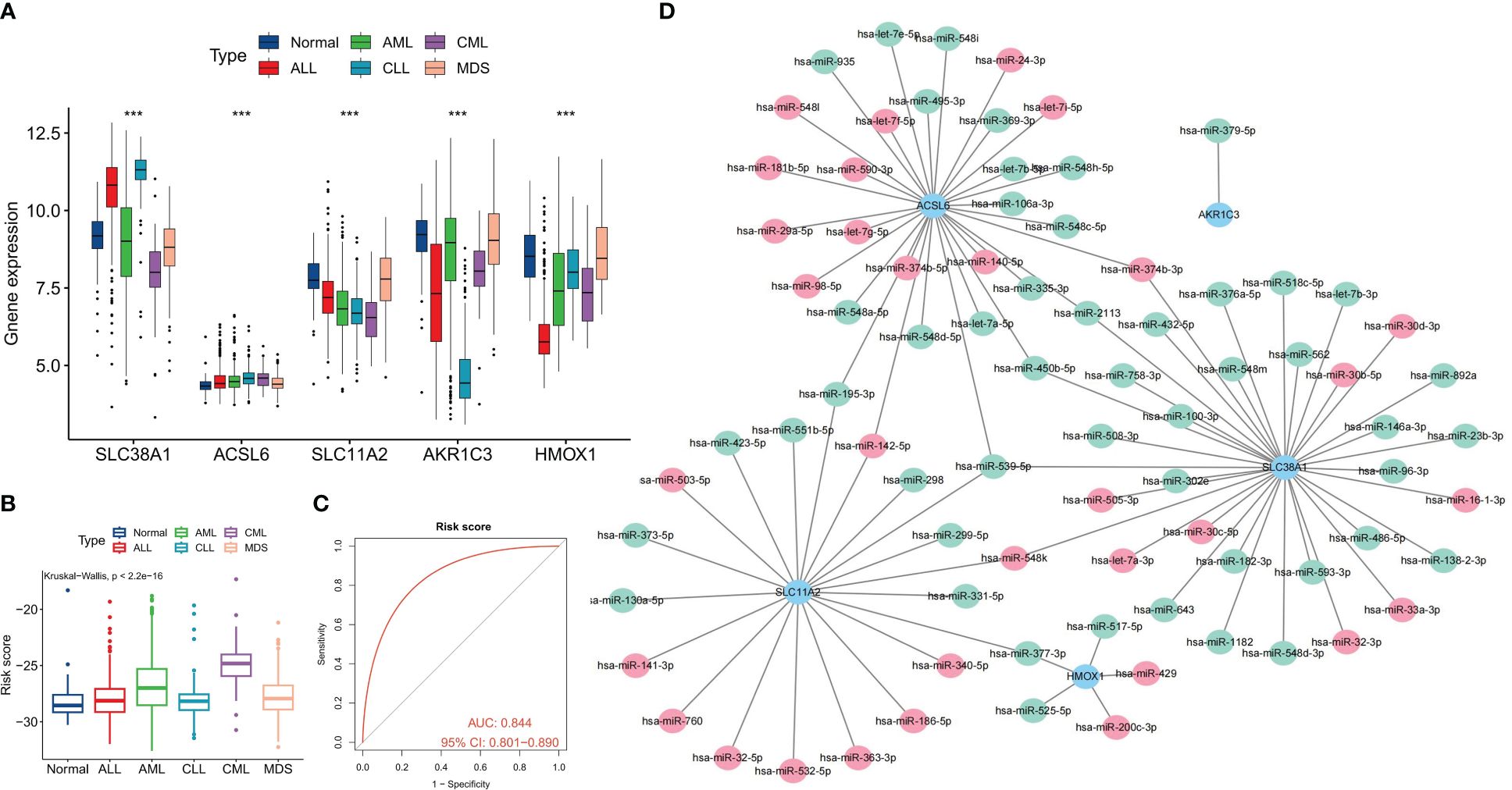
Determine 5 Differential diagnostic worth of the 5 FRGs in CML and different hematological malignancies. (A) Expression variations of the 5 diagnostic FRGs amongst CML, AML, CLL, ALL, MDS, and regular samples. (B) variations in danger scores amongst CML, AML, CLL, ALL, MDS, and regular samples. (C) ROC curve evaluation of danger scores in CML and different hematological malignancies. (D) Regulatory community of miRNAs and the 5 diagnostic FRGs; crimson signifies miRNA expression is up-regulated in CML samples, and inexperienced signifies expression is down-regulated. ***p < 0.001.
Identification of ferroptosis-related molecular subtypes and evaluation of variations in organic traits between subtypes
To comprehensively analyze the organic significance of FRGs in CML, we utilized the expression profiles of the 5 diagnostic FRGs in CML samples to establish two distinct molecular subtypes, particularly Cluster C1 and Cluster C2, using a consensus clustering algorithm (Determine 6A, Supplementary Desk S2). The distribution traits of those two molecular subtypes had been additional confirmed by PCA, revealing vital and discernible variations (Determine 6B). Subsequently, by way of heatmap visualization, it was noticed that ACSL6, SLC11A2, HMOX1, and AKR1C3 exhibited up-regulation in subtype C1 whereas SLC38A1 displayed increased expression ranges in subtype C2 (Determine 6C). To discover further distinctions between these subtypes at a organic degree, immune infiltration evaluation demonstrated that subtype C1 had an elevated proportion of CD8+ T cells, follicular helper T cells, activated dendritic T cells, and eosinophils in comparison with subtype C2 (Determine 6D). Moreover, there have been notable variations within the expression ranges of immune checkpoint genes; particularly inside subtype C1 the place PD-L1, CTLA-4, HAVCR2, PD-1, and CD80 confirmed elevated expressions (Determine 6E). This means that subtype C1 might exhibit sure immunosuppressive tendencies resulting in potential exhaustion of CD8+ T cells. These findings had been corroborated by increased TIDE scores for subtype C1 (Determine 6F). Conversely, C2subtype appeared extra more likely to profit from immunotherapy (Determine 6G).
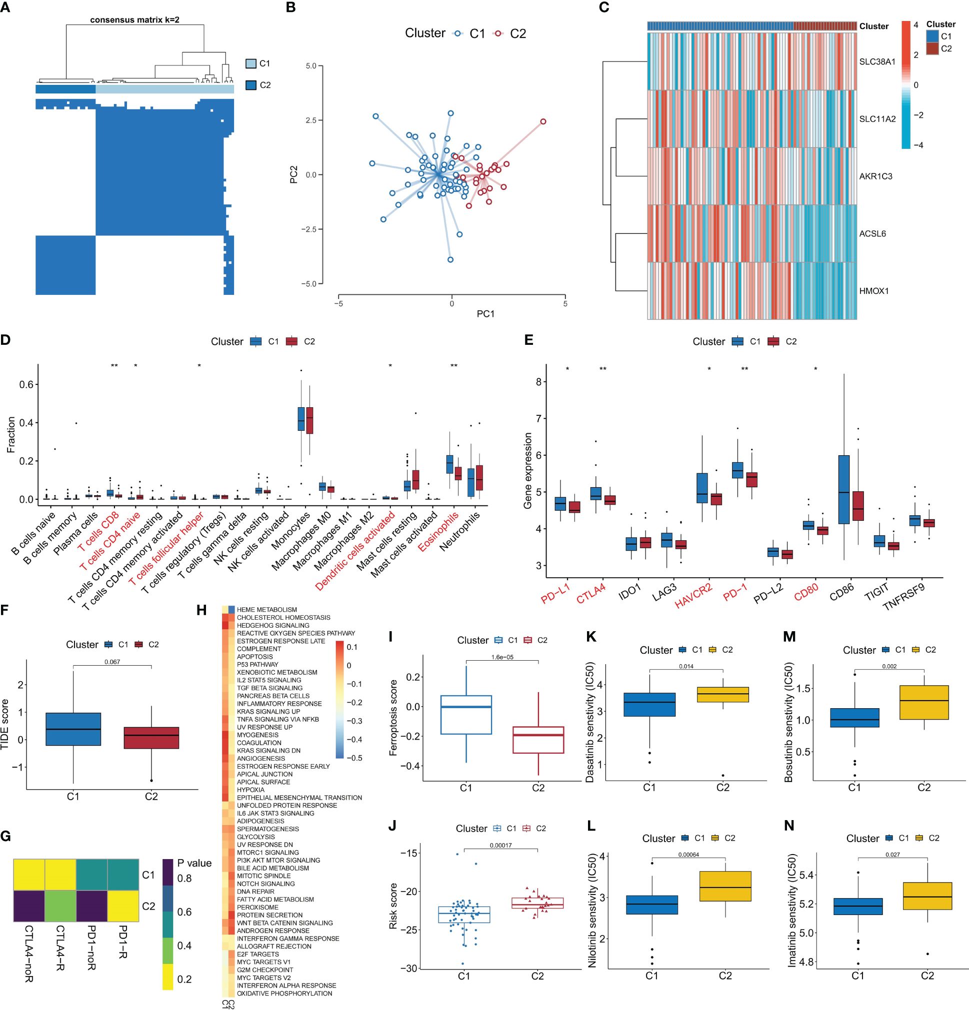
Determine 6 Identification of ferroptosis-related molecular subtypes and evaluation of their variations in organic traits and remedy sensitivity. (A) Primarily based on the expression of DEFRG, CML sufferers had been divided into two ferroptosis-related molecular subtypes by consensus clustering algorithm. (B) PCA algorithm was used to investigate the distribution variations of sufferers between subtypes. (C-F) Variations in expression of DEFRG (C), infiltration of twenty-two immune cells (D), expression of immune checkpoints (E), TIDE rating (F), immunotherapy response (G), exercise of tumor hallmark gene units (H), ferroptosis scores (I), danger rating (J), and therapeutic sensitivity to 4 TKIs (Okay-N) between the 2 molecular subtypes. *p < 0.05; **p < 0.01.
Moreover, our GSVA evaluation revealed that the C1 subtype demonstrates heightened activation of sign transduction pathways comparable to hedgehog signaling and TNFA signaling by way of NFKB (Determine 6H). Furthermore, we noticed elevated exercise in cancer-promoting pathways together with hypoxia and reactive oxygen species pathway. In distinction, the C2 subtype exhibited elevated exercise in proliferation-related pathways comparable to G2M checkpoint, E2F targets, and MYC targets V1. Notably, C1 displayed the next ferroptosis rating whereas C2 had the next danger rating (Figures 6I, J). Drug prediction evaluation indicated that imatinib, nilotinib, dasatinib, and bosutinib demonstrated larger efficacy in opposition to subtype C1 in comparison with subtype C2 (Figures 6K-N). These findings will considerably contribute to the event of personalised remedy methods for sufferers with CML.
In vitro experiments confirmed that CML-resistant cells had been extra delicate to ferroptosis remedy
The expression of 5 FRGs was detected in CML cell strains K562 and imatinib-resistant cell strains K562/IR. Compared to K562, SLC38A1 expression confirmed a slight up-regulation in K562/IR, whereas ACSL6, SLC11A2, and AKR1C3 expressions had been down-regulated (HMOX1 gene expression was not detected and subsequently not proven) (Determine 7A). In our research above, our preliminary evaluation indicated that CML cells might exhibit sensitivity to ferroptosis, whereas CML cells in blast disaster exhibit resistance in the direction of TKI remedy and probably increased sensitivity. To validate these findings, we performed in vitro experiments. Nonetheless, it was noticed that the CML cell line K562 didn’t show sensitivity to erastin-induced ferroptosis (Determine 7B); nonetheless, erastin exhibited a sure cytotoxic impact on imatinib-resistant K562 cells (K562/IR) with an IC50 of 5.099 μM (Determine 7C). Moreover, remedy of K562/IR cells with the ferroptosis inhibitor Fer-1 considerably restored mobile viability (Determine 7D). In comparison with K562 cells, there was a big improve in ROS ranges inside K562/IR cells which additional escalated after erastin treatment-indicating ROS as a vital issue for inducing ferroptosis (Determine 7E). Moreover, it was found that low-dose erastin enhanced the therapeutic sensitivity of imatinib in the direction of K562/IR cells by lowering the IC50 from 3.184 μM to 1.886 μM (Determine 7F). Furthermore, low-dose erastin promoted apoptosis ranges in K562/IR cells handled with imatinib (Figures 7G, H). GSH and GPX4 are vital indicators of ferroptosis. We discovered that after erastin remedy, GSH content material, GSH/GSSH ratio, and GPX4 enzyme exercise of K562/IR cells had been considerably decreased, and GPX4 mRNA expression degree was barely elevated (Figures 7I-L), indicating that erastin inhibited GSH manufacturing. In flip, the GPX4 enzyme exercise is lowered, which cannot inhibit the manufacturing of extra ROS, leading to ferroptosis of K562/IR cells. Lastly, we additionally detected GPX4 expression in K562 and K562/IR cells and CML samples, and the outcomes confirmed that GPX4 expression in K562/IR cells was decrease than that in K562 cells, and there was no vital distinction in GPX4 expression between BC-CML and CP-CML samples and regular samples (Figures 7M, N). These outcomes counsel that vital mechanisms of ferroptosis resistance in CML-resistant cells might not be regulated by GPX4.
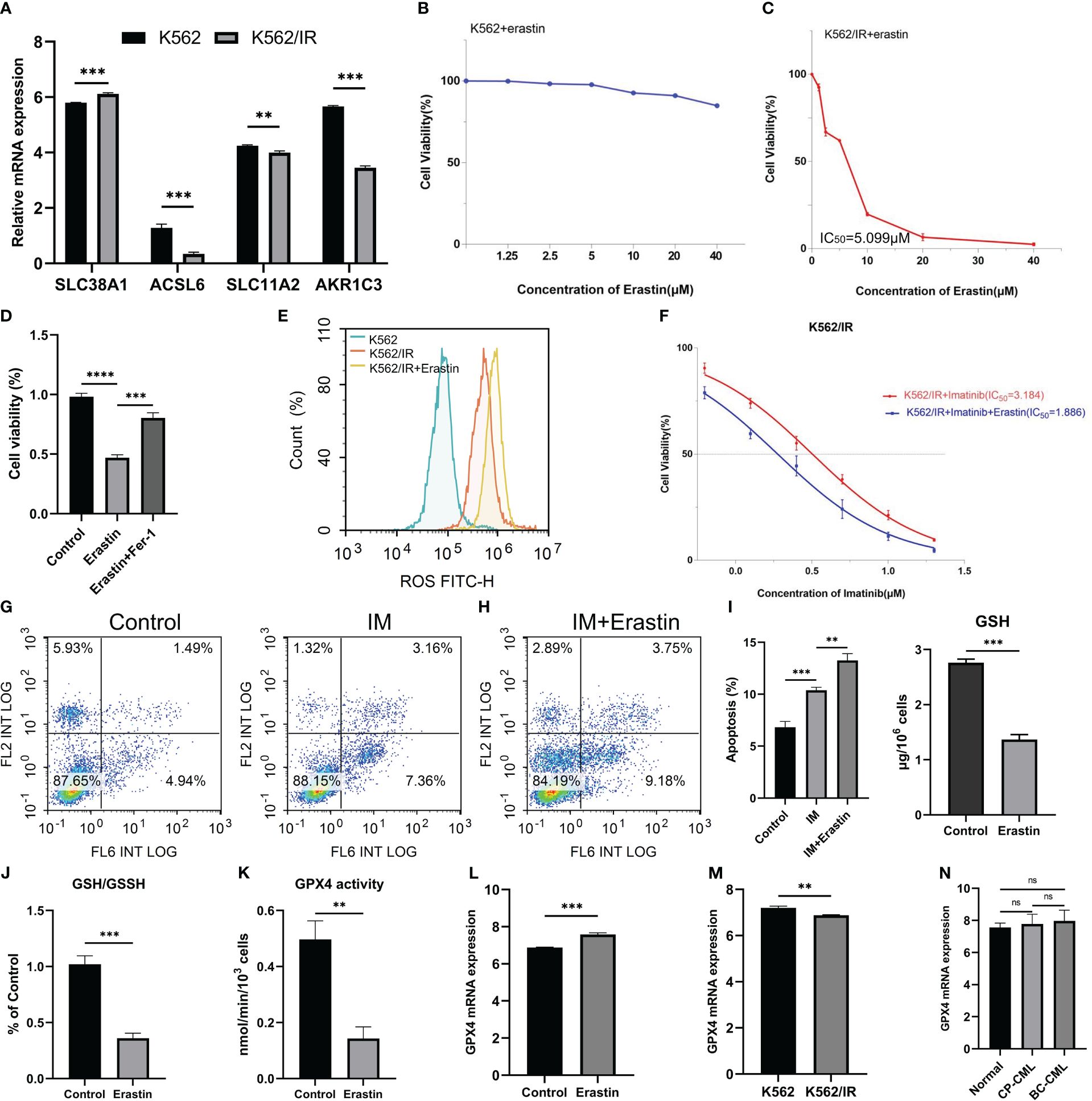
Determine 7 Therapeutic results of erastin on CML cells. (A) Evaluation of FRG expression between K562 and K562/IR cells. (B, C) Impact of various concentrations of erastin on cell viability of K562 and K562/IR cells after 48h remedy. (D) The exercise of K562/IR cells after remedy with 5 μM erastin and the addition of 1μM ferroptosis inhibitor Fer-1 for 48h. (E) ROS ranges in K562, K562/IR, and K562/IR had been handled with 5 μM erastin after 24h. (F) Modifications in cell viability with or with out 1.25 μM erastin and handled with completely different concentrations of imatinib for K562/IR after 48h remedy. (G, H) Modifications in apoptosis ranges after K562/IR remedy with or with out 1.25 μM erastin and 1 μM imatinib of 24h. (I-L) Modifications of GSH degree, GSH/GSSH ratio, GPX4 exercise, and GPX4 mRNA expression in K562/IR cells after 5 μM erastin remedy for 48h. (M, N) The distinction in GPX4 mRNA expression between K562 and K562/IR cells, in addition to amongst regular samples, CP-CML samples, and BC-CML samples. The IC50 worth of the drug was calculated by GraphPad software program. **p < 0.01; ***p < 0.001; ns, no significance.
Dialogue
Ferroptosis, a newly found mode of cell loss of life lately, performs a vital position in regulating varied physiological and pathological processes (10). Within the context of tumors, ferroptosis is intently related to the organic traits of tumor cells. The hypoxic microenvironment simply triggers the era of ROS, whereas the lipid metabolism required for fast proliferation creates favorable situations for lipid peroxidation (7). These options collectively point out that tumor cells are inclined to bear ferroptosis. The induction of ferroptosis in tumor cells and the attenuation of their protecting capability have vital scientific worth for most cancers remedy, aiming to reinforce tumor cell loss of life or develop novel focused therapies in opposition to apoptosis resistance (30).
On this research, we performed a scientific evaluation of ferroptosis ranges in samples from sufferers with CML utilizing transcriptome sequencing information. Our findings affirm the scientific significance of FRG in diagnosing and evaluating remedy outcomes for CML. Evaluation of information from a number of cohorts reveals a big discount in ferroptosis scores in CML samples, which additional decreases with illness development. Non-responders additionally exhibit decrease ferroptosis scores in comparison with CML sufferers who reply to TKI remedy. Subsequent analyses point out that decrease ferroptosis scores could also be related to decreased expression of genes concerned in suppressing ferroptosis, suggesting that CML cells with weaker inhibition in opposition to ferroptosis could also be extra prone to induction remedy concentrating on this course of. By means of further cell experiments, we validate that CML-resistant cells are extra delicate to the induction of ferroptosis and may improve the sensitivity of imatinib remedy, offering a novel goal and technique for overcoming drug resistance in CML. Moreover, our outcomes exhibit that the ferroptosis rating serves as an informative indicator reflecting the traits of the tumor microenvironment in CML. Sufferers with excessive ferroptosis scores exhibit elevated infiltration by Tregs and better expression ranges of immune checkpoint genes LAG3 and TNFRSF9, that are related to immunosuppression. Moreover, there’s a optimistic correlation between ferroptosis scores and exercise ranges inside most tumor signature pathways. By conducting WGCNA evaluation, now we have additional recognized metabolic pathways as essential determinants influencing the exercise of the ferroptosis pathway itself. Due to this fact, metabolic reprogramming performs a vital position not solely in selling malignant proliferation but additionally contributes to triggering ferroptosis (8, 29).
The expression profile and scientific significance of FRG had been additional analyzed on this research. Nearly all of differentially expressed FRGs had been discovered to be down-regulated in CML samples, suggesting their potential involvement within the pathogenesis of CML. Moreover, these FRGs had been discovered to take part in varied metabolic pathways, highlighting their multifaceted features past regulating ferroptosis. To comprehensively validate the diagnostic worth of FRG, three machine studying algorithms had been employed to establish 5 CML-specific diagnostic FRGs: ACSL6, SLC11A2, HMOX1, SLC38A1, and AKR1C3. These genes confirmed considerably lowered expression ranges in CML samples in comparison with regular samples.
The diagnostic worth of those 5 FRGs was confirmed not solely inside the evaluation cohort and validation cohort but additionally in a real-world scientific cohort. This complete validation enhanced the efficiency of the danger rating mannequin based mostly on their expression ranges for diagnosing CML sufferers precisely. Moreover, it was noticed that as remedy remission occurred in CML sufferers, the expression ranges of FRGs elevated whereas the danger scores decreased accordingly. Importantly, these 5 FRGs can be utilized for distinguishing CML from different hematological malignancies with scientific relevance. These bioinformatics findings present robust proof supporting the diagnostic and therapeutic analysis potential of FRG particularly in CML sufferers. Moreover, based mostly on distinct patterns of FRG expressions recognized by way of our evaluation strategy, we categorised two molecular subtypes inside the inhabitants of CML sufferers: subtype C1, characterised by the next proportion of CD8+ T cell infiltration and elevated immune checkpoint gene expressions suggesting immunosuppression; these sufferers are predicted to exhibit larger sensitivity in the direction of TKI therapies in comparison with subtype C2. In conclusion, the proposed molecular subtypes will considerably improve our understanding of the distinct illness traits exhibited by sufferers with CML, thereby offering invaluable insights for tailor-made scientific steerage in personalised remedy methods.
Lastly, we found by way of additional experimentation that CML-resistant cells exhibited heightened sensitivity to ferroptosis, probably as a result of elevated ranges of ROS in these cells. In tumor cells, ROS acts as a signaling molecule and promotes varied phenotypes comparable to development, metastasis, resistance to apoptosis, and differentiation problems by activating survival signaling pathways, accelerating power metabolism, and producing carcinogenic mutations (31). Quite a few research have additionally confirmed that ROS serves as a serious supply of genomic instability in several types of most cancers. The continual mutation of most cancers cell genomes is a big reason for drug resistance and relapse in most cancers remedy (32, 33). A number of research have additionally substantiated the explanations behind the substantial improve in ROS ranges noticed in CML-resistant cells. This primarily stems from the activation of assorted downstream signaling pathways by BCR-ABL1, together with the PI3K/AKT/mTOR pathway which reinforces glucose metabolism and mitochondrial electron transport chain exercise excessively (34, 35); augmentation of NADPH oxidase exercise (36); and regulation of goal gene transcription for ROS era by way of STAT5 (37). Accumulation of ROS drives a cycle of genomic instability resulting in BCR-ABL1 mutations or different chromosomal aberrations together with TKI resistance leading to drug resistance. Moreover, excessive ranges of ROS can induce oxidative harm to mitochondrial DNA inside CML-resistant cells inflicting mitochondrial dysfunction that disrupts the oxidative respiratory chain resulting in extreme electron leakage thereby additional growing ROS manufacturing inside resistant cells (38). Elevated ranges of ROS facilitate the formation of extra heteromutations whereas stimulating the signaling capability inside most cancers pathways thus producing further various mechanisms selling CML resistance. Due to this fact, elevated ranges of ROS play a pivotal position in rendering CML-resistant cells extra prone to ferroptosis, thereby providing a novel therapeutic avenue for overcoming CML resistance. At the moment, quite a few regulatory mechanisms related to ferroptosis have been elucidated, together with the involvement of HDAC3 by way of the Hippo signaling pathway (39). Additional exploration into the mechanism underlying ferroptosis in CML is warranted.
In abstract, now we have elucidated the molecular traits of ferroptosis in CML from a bioinformatics perspective. The findings from these analyses will contribute to a deeper understanding of the organic significance of ferroptosis in CML. FRG, recognized by way of varied machine studying algorithms and validated throughout a number of cohorts, demonstrates dependable scientific diagnostic worth. Furthermore, the introduction of ferroptosis-associated molecular subtypes has considerably enhanced our comprehension of individualized traits amongst CML sufferers and facilitated personalised remedy methods. The induction of ferroptosis may additionally function a promising therapeutic strategy for overcoming resistance in CML. Nonetheless, our research does have sure limitations, together with the necessity for a bigger pattern dimension to validate the bioinformatics findings, extra cell strains and extra complete experiments to elucidate the regulatory mechanisms underlying ferroptosis in CML-resistant cells. In subsequent research, we’ll develop our pattern assortment and improve our exploration of related mechanisms by way of each in vivo and in vitro experiments.
Conclusion
The transcriptomic evaluation performed on this research has revealed the molecular traits of ferroptosis in samples from sufferers with CML. By using machine studying algorithms, dependable scientific diagnostic worth was efficiently recognized for FRG expression patterns. This understanding of particular person molecular subtypes related to ferroptosis can successfully information scientific remedy methods for CML sufferers. Moreover, concentrating on and inducing ferroptosis exhibits nice promise as a possible therapeutic strategy to handle drug-resistant CML.
Information availability assertion
The unique contributions offered within the research are included within the article/Supplementary Materials. Additional inquiries may be directed to the corresponding authors.
Ethics assertion
The research involving people had been accepted by Ethics Committee of the Second Affiliated Hospital of Nanchang College. The research had been performed in accordance with the native laws and institutional necessities. The contributors offered their written knowledgeable consent to take part on this research.
Writer contributions
FZ: Information curation, Formal evaluation, Funding acquisition, Methodology, Sources, Software program, Validation, Visualization, Writing – unique draft. XZ: Validation, Visualization, Writing – unique draft. ZW: Validation, Visualization, Writing – unique draft. XL: Funding acquisition, Validation, Visualization, Writing – unique draft. BH: Funding acquisition, Validation, Visualization, Writing – unique draft. XW: Conceptualization, Funding acquisition, Mission administration, Sources, Supervision, Writing – evaluation & modifying. GK: Conceptualization, Funding acquisition, Mission administration, Sources, Supervision, Writing – evaluation & modifying.
Funding
The creator(s) declare monetary help was acquired for the analysis, authorship, and/or publication of this text. The research was funded by the Nationwide Pure Science Basis of China (82160405, 82160038, 82301578, 82170140 and 82370146), the Pure Science Basis of Jiangxi Province (20232BAB216037, 20232BAB216050), the Key Analysis and Improvement Program of Shaanxi Province (2024SF-YBXM-151), and the Shaanxi Basic Science Analysis Mission for Chemistry and Biology (Grant No. 23JHZ007).
Battle of curiosity
The authors declare that the analysis was performed within the absence of any industrial or monetary relationships that may very well be construed as a possible battle of curiosity.
Writer’s be aware
All claims expressed on this article are solely these of the authors and don’t essentially symbolize these of their affiliated organizations, or these of the writer, the editors and the reviewers. Any product which may be evaluated on this article, or declare which may be made by its producer, isn’t assured or endorsed by the writer.
Supplementary materials
The Supplementary Materials for this text may be discovered on-line at: https://www.frontiersin.org/articles/10.3389/fimmu.2024.1402669/full#supplementary-material
References
3. Poudel G, Tolland MG, Hughes TP, Pagani IS. Mechanisms of resistance and implications for remedy methods in persistent myeloid leukaemia. Cancers (Basel). (2022) 14. doi: 10.3390/cancers14143300
PubMed Summary | CrossRef Full Textual content | Google Scholar
4. Alves R, Gonçalves AC, Rutella S, Almeida AM, De Las Rivas J, Trougakos IP, et al. Resistance to tyrosine kinase inhibitors in persistent myeloid leukemia-from molecular mechanisms to scientific relevance. Cancers (Basel). (2021) 13. doi: 10.3390/cancers13194820
5. Rosti G, Castagnetti F, Gugliotta G, Baccarani M. Tyrosine kinase inhibitors in persistent myeloid leukaemia: which, when, for whom? Nat Rev Clin Oncol. (2017) 14:141–54. doi: 10.1038/nrclinonc.2016.139
PubMed Summary | CrossRef Full Textual content | Google Scholar
6. Ma L, Shan Y, Bai R, Xue L, Eide CA, Ou J, et al. A therapeutically targetable mechanism of BCR-ABL-independent imatinib resistance in persistent myeloid leukemia. Sci Trans Med. (2014) 6:252ra121. doi: 10.1126/scitranslmed.3009073
7. Mou Y, Wang J, Wu J, He D, Zhang C, Duan C, et al. Ferroptosis, a brand new type of cell loss of life: alternatives and challenges in most cancers. J Hematol Oncol. (2019) 12:34. doi: 10.1186/s13045-019-0720-y
PubMed Summary | CrossRef Full Textual content | Google Scholar
8. Stockwell B, Friedmann Angeli J, Bayir H, Bush A, Conrad M, Dixon S, et al. Ferroptosis: A regulated cell loss of life nexus linking metabolism, redox biology, and illness. Cell. (2017) 171:273–85. doi: 10.1016/j.cell.2017.09.021
PubMed Summary | CrossRef Full Textual content | Google Scholar
9. Dixon S, Lemberg Okay, Lamprecht M, Skouta R, Zaitsev E, Gleason C, et al. Ferroptosis: an iron-dependent type of nonapoptotic cell loss of life. Cell. (2012) 149:1060–72. doi: 10.1016/j.cell.2012.03.042
PubMed Summary | CrossRef Full Textual content | Google Scholar
11. Xie Y, Hou W, Tune X, Yu Y, Huang J, Solar X, et al. Ferroptosis: course of and performance. Cell Demise differentiat. (2016) 23:369–79. doi: 10.1038/cdd.2015.158
12. Yang W, SriRamaratnam R, Welsch M, Shimada Okay, Skouta R, Viswanathan V, et al. Regulation of ferroptotic most cancers cell loss of life by GPX4. Cell. (2014) 156:317–31. doi: 10.1016/j.cell.2013.12.010
PubMed Summary | CrossRef Full Textual content | Google Scholar
13. Bersuker Okay, Hendricks J, Li Z, Magtanong L, Ford B, Tang P, et al. The CoQ oxidoreductase FSP1 acts parallel to GPX4 to inhibit ferroptosis. Nature. (2019) 575:688–92. doi: 10.1038/s41586–019-1705–2
PubMed Summary | CrossRef Full Textual content | Google Scholar
14. Hu Q, Wei W, Wu D, Huang F, Li M, Li W, et al. Blockade of GCH1/BH4 axis prompts ferritinophagy to mitigate the resistance of colorectal most cancers to erastin-induced ferroptosis. Entrance Cell Dev Biol. (2022) 10:810327. doi: 10.3389/fcell.2022.810327
PubMed Summary | CrossRef Full Textual content | Google Scholar
15. Wu J, Minikes AM, Gao M, Bian H, Li Y, Stockwell BR, et al. Intercellular interplay dictates most cancers cell ferroptosis by way of NF2-YAP signalling. Nature. (2019) 572:402–6. doi: 10.1038/s41586–019-1426–6
PubMed Summary | CrossRef Full Textual content | Google Scholar
16. Lee H, Zandkarimi F, Zhang Y, Meena JK, Kim J, Zhuang L, et al. Power-stress-mediated AMPK activation inhibits ferroptosis. Nat Cell Biol. (2020) 22:225–34. doi: 10.1038/s41556–020-0461–8
PubMed Summary | CrossRef Full Textual content | Google Scholar
17. Zou Y, Palte MJ, Deik AA, Li H, Eaton JK, Wang W, et al. A GPX4-dependent most cancers cell state underlies the clear-cell morphology and confers sensitivity to ferroptosis. Nat Commun. (2019) 10:1617. doi: 10.1038/s41467–019-09277–9
PubMed Summary | CrossRef Full Textual content | Google Scholar
18. Yusuf RZ, Saez B, Sharda A, van Gastel N, Yu VWC, Baryawno N, et al. Aldehyde dehydrogenase 3a2 protects AML cells from oxidative loss of life and the artificial lethality of ferroptosis inducers. Blood. (2020) 136:1303–16. doi: 10.1182/blood.2019001808
PubMed Summary | CrossRef Full Textual content | Google Scholar
19. Du J, Wang T, Li Y, Zhou Y, Wang X, Yu X, et al. DHA inhibits proliferation and induces ferroptosis of leukemia cells by way of autophagy dependent degradation of ferritin. Free Radical Biol Med. (2019) 131:356–69. doi: 10.1016/j.freeradbiomed.2018.12.011
20. Zhong FM, Yao FY, Liu J, Zhang HB, Zhang J, Zhang N, et al. Ferroptosis-related molecular patterns reveal immune escape, inflammatory growth and lipid metabolism traits of the tumor microenvironment in acute myeloid leukemia. Entrance Oncol. (2022) 12:888570. doi: 10.3389/fonc.2022.888570
PubMed Summary | CrossRef Full Textual content | Google Scholar
21. Liu S, Wu W, Chen Q, Zheng Z, Jiang X, Xue Y, et al. TXNRD1: A key regulator concerned within the ferroptosis of CML cells induced by cysteine depletion in vitro. Oxid Med Cell Longev. (2021) 2021:7674565. doi: 10.1155/2021/7674565
PubMed Summary | CrossRef Full Textual content | Google Scholar
22. Yu G, Wang LG, Han Y, He QY. clusterProfiler: an R package deal for evaluating organic themes amongst gene clusters. Omics: J Integr Biol. (2012) 16:284–7. doi: 10.1089/omi.2011.0118
23. Hänzelmann S, Castelo R, Guinney J. GSVA: gene set variation evaluation for microarray and RNA-seq information. BMC Bioinf. (2013) 14:7. doi: 10.1186/1471–2105-14–7
24. Newman A, Liu C, Inexperienced M, Gentles A, Feng W, Xu Y, et al. Sturdy enumeration of cell subsets from tissue expression profiles. Nat Strategies. (2015) 12:453–7. doi: 10.1038/nmeth.3337
PubMed Summary | CrossRef Full Textual content | Google Scholar
25. Langfelder P, Horvath S. WGCNA: an R package deal for weighted correlation community evaluation. BMC Bioinf. (2008) 9:559. doi: 10.1186/1471–2105-9–559
26. Wilkerson M, Hayes D. ConsensusClusterPlus: a category discovery device with confidence assessments and merchandise monitoring. Bioinf (Oxford England). (2010) 26:1572–3. doi: 10.1093/bioinformatics/btq170
27. Geeleher P, Cox N, Huang R. pRRophetic: an R package deal for prediction of scientific chemotherapeutic response from tumor gene expression ranges. PloS One. (2014) 9:e107468. doi: 10.1371/journal.pone.0107468
PubMed Summary | CrossRef Full Textual content | Google Scholar
28. Li SQ, Liu J, Zhang J, Wang XL, Chen D, Wang Y, et al. Transcriptome profiling reveals the excessive incidence of hnRNPA1 exon 8 inclusion in persistent myeloid leukemia. J superior Res. (2020) 24:301–10. doi: 10.1016/j.jare.2020.04.016
33. Cui Q, Wang JQ, Assaraf YG, Ren L, Gupta P, Wei L, et al. Modulating ROS to beat multidrug resistance in most cancers. Drug resistance updates: Rev commentaries antimicrobial Anticancer chemother. (2018) 41:1–25. doi: 10.1016/j.drup.2018.11.001
34. Antoszewska-Smith J, Pawlowska E, Blasiak J. Reactive oxygen species in BCR-ABL1-expressing cells – relevance to persistent myeloid leukemia. Acta Biochim Polonica. (2017) 64:1–10. doi: 10.18388/abp.2016_1396
35. Kim JH, Chu SC, Gramlich JL, Pleasure YB, Babendreier E, Chauhan D, et al. Activation of the PI3K/mTOR pathway by BCR-ABL contributes to elevated manufacturing of reactive oxygen species. Blood. (2005) 105:1717–23. doi: 10.1182/blood-2004–03-0849
PubMed Summary | CrossRef Full Textual content | Google Scholar
36. Reddy MM, Fernandes MS, Salgia R, Levine RL, Griffin JD, Sattler M. NADPH oxidases regulate cell development and migration in myeloid cells reworked by oncogenic tyrosine kinases. Leukemia. (2011) 25:281–9. doi: 10.1038/leu.2010.263
PubMed Summary | CrossRef Full Textual content | Google Scholar
37. Warsch W, Grundschober E, Berger A, Gille L, Cerny-Reiterer S, Tigan AS, et al. STAT5 triggers BCR-ABL1 mutation by mediating ROS manufacturing in persistent myeloid leukaemia. Oncotarget. (2012) 3:1669–87. doi: 10.18632/oncotarget.806
PubMed Summary | CrossRef Full Textual content | Google Scholar
38. Glowacki S, Synowiec E, Blasiak J. The position of mitochondrial DNA harm and restore within the resistance of BCR/ABL-expressing cells to tyrosine kinase inhibitors. Int J Mol Sci. (2013) 14:16348–64. doi: 10.3390/ijms140816348
PubMed Summary | CrossRef Full Textual content | Google Scholar