IER5L is upregulated in most cancers
To investigate the affiliation of IER relations (IER2, IER5 and IER5L) with tumor pathogenesis and development, we interrogated public transcriptomics datasets containing gene expression information from totally different tumor varieties (Supplementary Desk S1) utilizing Cancertool [14]. IER2 and IER5 expression was altered in a few of the prostate, colorectal and lung datasets analyzed, however didn’t present a directional consistency throughout tumor varieties (Supplementary Fig. S1). Against this, IER5L was persistently upregulated in colorectal (2/2 datasets), lung (1/1 datasets) and prostate (2/4 datasets) tumor specimens when in comparison with non-cancerous tissue (Fig. 1a), in keeping with current studies [15, 16]. To additional discover whether or not the rise in IER5L ranges was a typical function of various tumor varieties, we took benefit of TIMER internet interface [17], which allowed us to discover the cancer-associated alterations within the TCGA cohorts. IER5L ranges persistently elevated throughout most cancers varieties (with an identical non-significant pattern in PCa), whereas IER2 expression was largely lowered, and IER5 exhibited inconsistent alterations (Supplementary Fig. S2).
a Violin plots depicting the Log2 expression of IER5L in non-tumoral (N), prostate most cancers (PCa), lung adenocarcinoma (LUAD) and colorectal most cancers (CRC) specimens within the indicated dataset. p-value derives from a Pupil’s t-test evaluation between the indicated teams. b Violin plots displaying the Log2 expression of IER5L mRNA in main tumor (PT) and metastatic (M) PCa specimens. p-value derives from a Pupil’s t-test evaluation between the indicated teams. c Kaplan-Meyer curves displaying the affiliation of IER5L mRNA expression to disease-free survival (DFS) within the indicated datasets. Quartile 1 (blue) and quartile 4 (inexperienced) are represented. A log-rank check p-value and the hazard ratio (HR) between two teams calculated by Cox proportional hazard mannequin regression are offered above every graph.
Not like different datasets, the PCa datasets included in Cancertool include the gene expression info of metastatic samples. To decipher whether or not the adjustments within the expression of IER household genes had been emphasised in metastatic specimens, we in contrast the gene expression of 56 metastatic samples and 200 main tumors. Remarkably, IER5L was elevated in metastatic specimens in 3 out of 4 of the interrogated prostate datasets (Fig. 1b), whereas IER2 expression was predominantly lowered, and IER5 exhibited no constant sample (Supplementary Fig. S3).
Subsequent, profiting from the medical follow-up info in numerous datasets, we interrogated the prognostic potential of the IER gene household via disease-free survival. Curiously, IER5L exhibited probably the most distinguished prognostic capability when upregulated among the many three family members (Fig. 1c, Supplementary Fig. S4). Making an allowance for the excessive consistency of IER5L upregulation within the numerous pathogenic situations analyzed, and the restricted details about its function in most cancers, we determined to review the organic and molecular operate of IER5L utilizing PCa as a tumor sort the place its alteration might be related.
Silencing of IER5L compromises the foci formation, migration and invasion capacity of PCa cells
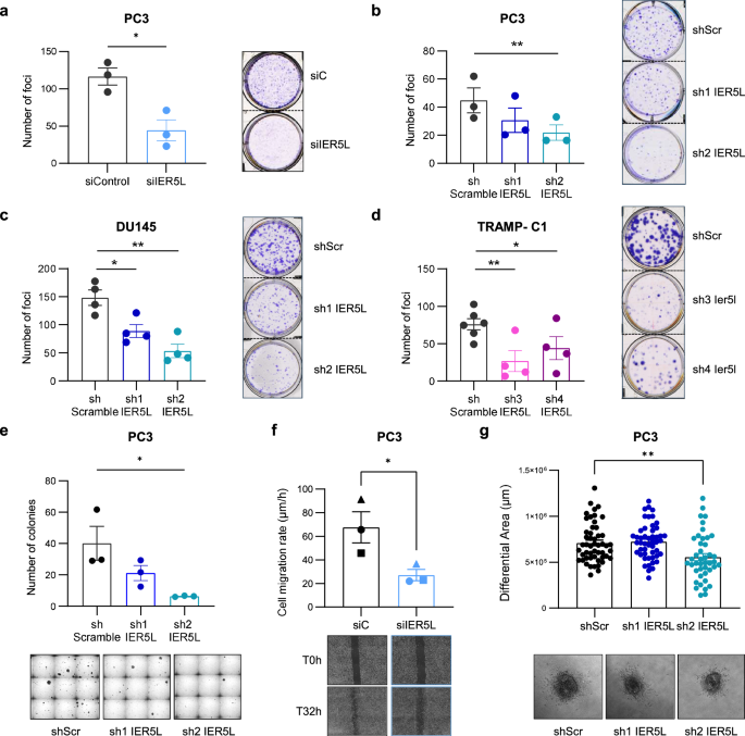
To check the function of IER5L in PCa aggressiveness, we analyzed the organic penalties of IER5L silencing in vitro. A lower in IER5L mRNA ranges by both small interference RNA (siRNA) transfection or quick hairpin (shRNA) transduction didn’t compromise the general cell progress of PC3 cells (Supplementary Fig. S5a, b). In distinction, when cells with decreased IER5L ranges had been pressured to develop individualized, they confirmed a decrease functionality to type colonies in foci formation assays (Fig. 2a, b). These outcomes had been replicated in two extra human PCa cell strains (DU145 and 22RV1) and a murine PCa cell line, TRAMP-C1 (Fig. 2c, d, Supplementary Fig. S5c-f). Likewise, IER5L silencing additionally impaired the flexibility of human and murine PCa cells to develop in an anchorage-independent method (Fig. 2e, Supplementary Fig. S5g). Of observe, the silencing efficacy of the 2 human IER5L-targetting shRNAs was related to the proportional tumor suppressive phenotype (Fig. 2, Supplementary Fig. S5). Furthermore, in keeping with the important function of IER5L in sustaining cell proliferation, its silencing was progressively misplaced with passages (Supplementary Fig. S5h), suggestive of the damaging number of silenced cells in vitro and stopping us from producing CRISPR/Cas9-based IER5L knockout cells.
a Foci-formation evaluation of PC3 cells transfected with the indicated siRNA. The variety of foci is proven (left panels). Consultant photos are proven (proper panels). A t-test was utilized for statistical evaluation (n = 3). siC: non-target siRNA. b–d Evaluation of foci formation upon IER5L depletion with the indicated shRNAs. The variety of foci is proven (left panels). Consultant photos are proven (proper panels). A paired t-test was utilized for statistical evaluation (b n = 3; c, d n = 4). shScr: shScramble. e Evaluation of anchorage-independent progress of PC3 cells transduced with the indicated shRNAs. The variety of colonies is proven (left panel). Consultant photos are proven (proper panel). A paired t-test was utilized for statistical evaluation (n = 3). f Cell migration charge of PC3 cells transfected with the indicated pool of siRNAs. The totally different organic replicates are indicated with distinctive dot shapes (prime panel). Consultant photos of the scratch at time (T) 0 and 32 hours (h) are proven (backside panel). Two days after siRNA transfection had been outlined because the preliminary timepoint for the assay. A two-tailed paired t-test was utilized for statistical evaluation (n = 3). g Quantification of the invasive progress of PC3 cells embedded in collagen. The diameter of the spheroids was measured at 0 and 72-h and the differential space was calculated (prime panel). Consultant photos of the spheroids at ultimate timepoint are proven (backside panel). A two-tailed unpaired t-test was utilized for statistical evaluation (n = 4).
Moreover the flexibility to develop below stress situations, metastatic cells generally purchase migration and invasion talents that contribute to their metastatic potential. Thus, we ascertained the contribution of IER5L to those processes in PC3 cells. On one hand, IER5L-targeting siRNA transfection in PCa cells decreased their migration charge in wound therapeutic assays (Fig. 2f). Then again, IER5L silencing lowered the invasive capability of PC3 cells in spheroid assays utilizing a collagen matrix, which was vital in cells with the best IER5L silencing (Fig. 2g, Supplementary Fig. S5b). Altogether, these information recommend that IER5L sustains aggressiveness properties in PCa.
IER5L contributes to tumor progress and metastasis in vivo
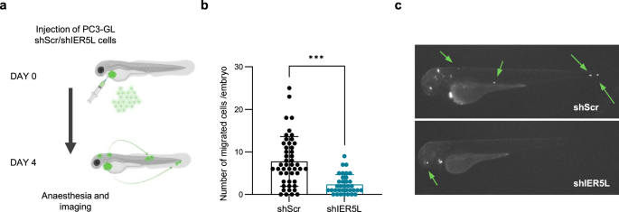
Our bioinformatics evaluation revealed an affiliation of IER5L upregulation to most cancers development (Fig. 1). As well as, in vitro assays confirmed a requirement of IER5L for cell progress below stress, migration and invasion. In flip, we designed in vivo methods that enabled us to watch all these totally different parameters. As a primary method, we exploited zebrafish mannequin to judge the causal contribution of IER5L to PCa cell dissemination in vivo. The transparency of the younger embryos, their lack of immune system and the speedy capability to type main tumor and metastases, makes zebrafish an acceptable and engaging mannequin for investigating tumor cell dissemination from the first website [18]. PC3 cells stably expressing GFP-luciferase (GL) transduced with both shScramble or shIER5L had been injected into the pericardial cavity of zebrafish embryos. The variety of disseminated cells was analyzed at 4 days put up injection (Fig. 3a). As proven in Fig. 3b, c, the silencing of IER5L considerably decreased the dissemination capacity of PC3 cells on this mannequin.
a Illustration of the experimental design of the in vivo cell dissemination assay in zebrafish. PC3 GFP-Luc (GL) cells transduced with shScramble (shScr) or sh2 IER5L (shIER5L) had been injected into the pericardial cavity of zebrafish embryos. Cell dissemination was analyzed at 4 days put up injection. The variety of disseminated cells (b) and a consultant picture (c) are proven. A two-tailed Mann–Whitney check was utilized for statistical evaluation.
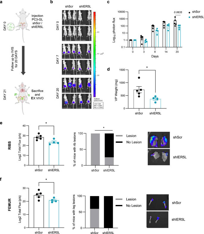
To additional validate the causal contribution of IER5L to PCa cell progress and dissemination, we carried out an in vivo orthotopic assay the place PC3-GL cells had been transduced with Scramble or IER5L-targeting shRNA (Fig. 4a). Luciferase-expressing cells had been then injected into the ventral lobe of immune-deficient nude mice and tumor mass was monitored by in vivo imaging for 20 days. IER5L-silenced cells confirmed a decrease luciferase sign all through the experiment (Fig. 4b, c), which correlated with a lowered main tumor weight on the experimental endpoint (Fig. 4d). Importantly, the evaluation of luciferase sign in distal organs confirmed a decrease metastatic burden in mice injected with IER5L-silenced PCa cells. This phenotype was extra distinguished in bones, together with ribs and femur (Fig. 4e, f, Supplementary Fig. S6).
a Experimental design of the in vivo orthotopic assay. PC3 GFP-LUC (GL) cells transduced with shScramble (shScr) or sh2 IER5L (shIER5L) had been injected into the ventral prostate lobe of nude mice and adopted up for 20 days. IVIS relative flux information alongside the experimental course of. Consultant photos (b) and the entire photon flux normalized to time 0 (c) are represented. A a number of Mann-Whitney U-test was utilized for statistical evaluation. d Ex vivo tumor weight of the ventral lobes of prostates (VP) from the in vivo orthotopic assay. A two-tailed Mann-Whitney check was utilized for statistical evaluation. Ex vivo IVIS sign quantification of ribs (e) and femur (f) (left panels). Contingency evaluation of metastatic lesions at these websites (center panels) and a consultant picture (proper panels) are proven. A one-tailed Mann–Whitney check and a Fisher precise t-test had been used, respectively.
Altogether, these information underline the significance of IER5L in sustaining metastatic properties in PCa cells.
IER5L silencing elicits molecular alterations in keeping with PP2A inhibition
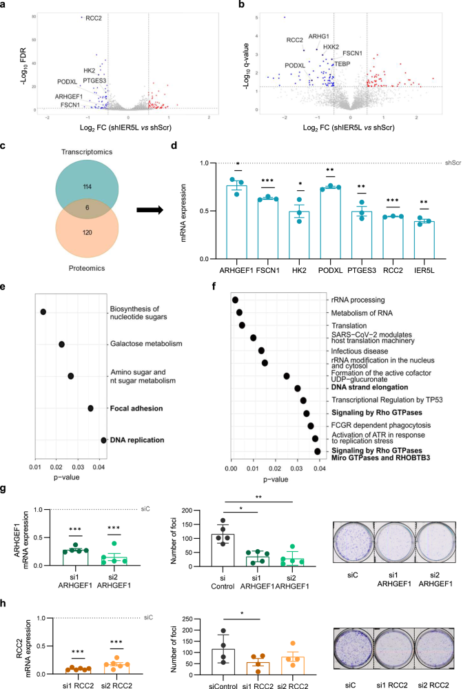
To achieve perception into the molecular mechanism by which IER5L contributes to the acquisition of aggressive options, we studied transcriptome and proteome alterations elicited upon IER5L silencing. PC3 cells transduced with IER5L-targeting shRNA exhibited sturdy adjustments within the transcriptional panorama (GSE249359, Supplementary Desk S2, Supplementary Fig. S7a). By evaluating PC3 cells transduced with both shScramble or shIER5L lentivirus, we recognized 120 differentially expressed genes (DEGs) (Fig. 5a). Of these, 63 had been upregulated and 57 downregulated in IER5L-silenced cells. IER5L silencing additionally altered the degrees of 126 proteins, 59 of which elevated whereas 67 decreased upon silencing (Fig. 5b, Supplementary Desk S3, Supplementary Fig. S7b). Of observe, the mixed evaluation of transcriptomics and proteomics revealed 6 candidate genes that had been persistently downregulated on the mRNA and protein degree (Fig. 5c). The alteration in these 6 genes upon IER5L silencing was validated in impartial pattern units (Fig. 5d, Supplementary Fig. S7c), the place the totally different silencing efficacy of IER5L-targeting shRNAs was mirrored in a milder impact on the expression of the genes evaluated. Furthermore, the downregulation of the targets was validated utilizing a second cell line, 22RV1 (Supplementary Fig. S7f). A lower on HK2, PTGES3 and RCC2 was noticed on this PCa cell line upon IER5L silencing.
Volcano plot illustration of the differentially expressed genes (DEGs) (a) and proteins (b) upon IER5L silencing by shScramble (shScr) or sh2 IER5L (shIER5L) transduction. The widespread targets between the RNAseq and proteomics’ analyses are highlighted. c Venn diagram summarizing the variety of DEGs and the proteins affected by IER5L depletion within the RNAseq and proteomics experiments from (a) and (b). d Evaluation of the expression of the indicated genes by qRT-PCR upon IER5L depletion by sh2 IER5L transduction in PC3 cells. The mRNA ranges are normalized to GAPDH and shScr. The dotted line represents the normalized worth of the shScr information. A one-sample t-test was utilized for statistical evaluation (n = 3). Useful enrichment evaluation of the DEGs upon IER5L silencing by KEGG (e) and Reactome (f). Left panels: Evaluation of ARHGEF1 (g) and RCC2 (h) mRNA expression by qRT-PCR. The degrees are normalized to GAPDH and non-target (siC) situation. The dotted line represents the normalized worth of the siC information. A one-sample t-test was utilized for statistical evaluation (n = 5 and n = 6 respectively). Center panels: Evaluation of foci formation upon ARHGEF1 (g) and RCC2 (h) depletion. The variety of foci is proven. A paired t-test was utilized for statistical evaluation (n = 5 and n = 4 respectively). Proper panels: Consultant photos of the foci experiments are proven.
Useful enrichment evaluation of the molecular alterations elicited upon IER5L silencing in PC3 cells uncovered adjustments in DNA replication and monomeric G protein exercise (Fig. 5e, f), that are in keeping with the discount in cell progress and motility noticed in our cell biology assays. Curiously, Regulator of Chromosome Condensation 2 (RCC2) and Rho Guanine Nucleotide Alternate Issue 1 (ARHGEF1), two of the highest candidate genes downregulated upon IER5L silencing (Fig. 5c), are regulators of the aforementioned processes [19,20,21]. Subsequently, we studied whether or not the downregulation of those elements might be a contributing occasion to the phenotype of IER5L silencing. To this finish, we silenced independently RCC2 and ARHGEF1 with 2 impartial siRNAs and evaluated the organic penalties (Fig. 5g, h). According to the implications of IER5L silencing, siRNA-mediated concentrating on of RCC2 or ARHGEF1 lowered foci-formation however not common two-dimensional progress (Fig. 5g, h, Supplementary Fig. S7e, f). These outcomes recommend that the transcriptional program elicited upon IER5L concentrating on is a key contributing occasion for the tumor suppressive phenotype.
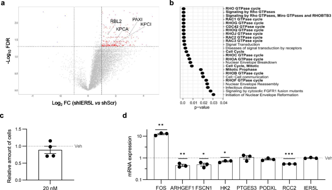
The data relating to the mechanism of motion of the IER household genes factors to the regulation of PP2A, whose operate and targets are not less than partly modulated by IER relations [5,6,7,8]. To check the relevance of this phosphatase within the motion of IER5L, we carried out two impartial approaches. On one hand, we analyzed the adjustments in phosphopeptides elicited upon IER5L silencing utilizing label-free proteomics. In keeping with the function of IER5L sustaining PP2A exercise, we noticed a sturdy enhance in phosphopeptides upon silencing of this gene in PC3 cells (Fig. 6a, Supplementary Desk S4). The inference of the proteins whose phosphorylation standing modified upon IER5L silencing recognized reported PP2A targets equivalent to RB Transcriptional Corepressor Like 2 (RBL2) [22, 23], Protein Kinase C Iota (KPCI) [24], Protein Kinase C Alpha (KPCA) [25] and Paxilin (PAXI) [26] (Fig. 6a). Curiously, the purposeful enrichment evaluation of these proteins highlighted processes associated to the organic and molecular penalties of IER5L silencing (cell cycle and monomeric G protein exercise) (Fig. 6b). Then again, we evaluated whether or not pharmacological inhibition of PP2A utilizing okadaic acid [27,28,29,30] would mimic the transcriptional alterations elicited upon IER5L silencing. Upregulation of Fos Proto-Oncogene (FOS) upon okadaic acid remedy [31] corroborated the exercise of this compound at doses that didn’t elicit cytotoxicity (Fig. 6c, d). Importantly, PP2A inhibition lowered the expression of IER5L-regulated genes, together with RCC2 and ARGHEF1, with out altering IER5L ranges (Fig. 6d). In keeping with the lower on RCC2 noticed upon IER5L silencing on 22RV1 cells (Supplementary Fig. S7d), PP2A inhibition on this cell line recapitulated the downregulation impact on RCC2 at doses that induced no defect in cell viability (Supplementary Fig. S8). Total, our outcomes are in keeping with organic and transcriptional results of IER5L which are related to the reported regulation of PP2A.
a Volcano plot illustration of the phosphopeptides altered upon IER5L silencing by shScramble (shScr) or sh2 IER5L (shIER5L) transduction. The proteins inferred from the altered phosphopeptides which are reported targets of PP2A are highlighted. b Useful enrichment evaluation of the proteins inferred from the differentially phosphorylated peptides upon IER5L silencing by Reactome. c Evaluation of crystal violet staining after a 24-h remedy with 20 nM okadaic acid. The absorbance was normalized to Car (Veh). A one-sample t-test was utilized for statistical evaluation (n = 4). d Evaluation of the expression of the indicated genes by qRT-PCR upon a 24-h remedy with 20 nM okadaic acid. The degrees are normalized to GAPDH and Car (Veh). The dotted line represents the normalized worth of the Veh information. A one-sample t-test was utilized for statistical evaluation (n = 3).


