Most cancers related fibroblasts constituted the vital a part of TME
The tumor microenvironment (TME) contains varied practical subgroups of each immune and non-immune cells. The intricate regulatory interactions inside this surroundings are primarily orchestrated by mediators discovered within the cluster of cancer-associated fibroblasts (CAFs). To determine the presence and practical roles of CAFs, we initially carried out examinations of stromal cell staining and analyzed single-cell RNA (scRNA) datasets. Our investigations revealed that fibroblasts are a distinguished part of lung tissues (see Fig. S1A). Additional scrutiny demonstrated that CAFs characterize a major proportion of each lung adenocarcinoma (LUAD, Fig. S1B) and lung squamous carcinoma (LUSC, Fig. S1C). Our validation prolonged to single-cell evaluation databases, the place we noticed plentiful enrichment of fibroblasts in GSE85716 (Fig. S1D), GSE118370 (Fig. S1E), and in two TCGA lung most cancers datasets (Fig. S1F,G). The additional deciphering of GSE85716 dataset proved that though totally different analyzing modes correlates with totally different CAF ratios, CAF was at all times the vital and foremost part (Fig. S2).
Outline the most cancers related fibroblasts with simplest marker
Fibroblasts represent a pivotal aspect throughout the TME teams, and quite a few signature markers have been recognized and partially validated in each human (Fig. 1A) and mouse (Fig. 1B) fashions. Amongst these markers, COL1A1 and PDGFRA have emerged as probably the most well known and universally accepted, demonstrating the best specificity (Fig. 1C). A plot map consisting all fibroblasts markers are enrolled to check their expressing patterns, extra particularly, COL1A1, COL1A2, and FAP confirmed finest correlation (Fig. 1D), and fibroblasts, myofibroblasts included, confirmed shut reference to other forms of TME subgroups (Fig. 1E, Fig. S3A,B).
The markers largely used to outline the subgroup of TME fibroblast. By means of screening and check on web site of “Floor markers”, probably the most accepted and universally used fibroblasts in human (A) and in mouse (B) had been displayed. (C) The markers of COL1A1 and PDGFRA confirmed the most effective specificity. (D) Plotting outcomes indicated that COL1A1, COL1A2, and FAP confirmed finest illustration. (E) Fibroblasts confirmed shut connections with other forms of TME subgroups in GSE lung most cancers samples, and every quantity indicated one particular subgroup as was labeled (uncooked information might be achieved at http://117.50.127.228/CellMarker/CellMarker_communication.jsp).
To elucidate the distinct traits of every potential CAF marker inside all TME teams, we meticulously examined single-cell RNA sequencing information from varied human organs and techniques on the Tubular Institute. This evaluation unveiled numerous distribution patterns amongst totally different TME subgroups (see Fig. S4A). Upon nearer examination, it grew to become evident that many fibroblast markers exhibited restricted specificity in figuring out distinctive subgroups (Fig. S4B, C). Moreover, common markers similar to COL1A1, ITGB1, PDGFRA, PDGFRB, FAP, SOX4, NOTCH3, KLF4, ACTA2, and S100A4 displayed both restricted or non-specific distribution within the complete organ-wide evaluation. Nevertheless, we noticed that ITGB1, COL1A1, and S100A4 exhibited excessive sensitivity, whereas COL1A1, PDGFRA, PDGFRB, and FAP demonstrated distinctive specificity (Fig. S4D, E).
Expression profiling of CAF markers in lung most cancers stromal cell teams
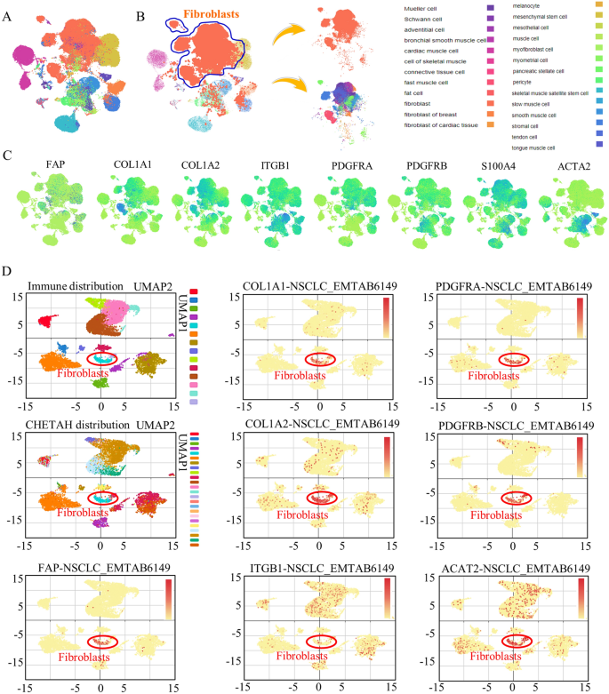
In a extra detailed examination, we centered on fibroblasts derived from varied human tissues. Our investigation established that almost all of fibroblasts are certainly among the many stromal cell inhabitants (see Fig. 2A, B). We additional verified the sensitivity and specificity of FAP, COL1A1, and COL1A2 in figuring out fibroblasts. The considerably heightened expression of those markers unequivocally correlated with the proportional illustration of each fibroblasts and cancer-associated fibroblasts (CAFs) (Fig. 2C). Additional in lung most cancers tissues, via scRNA evaluation at “IMMUcan SingleCell RNAseq-Database” of lung most cancers, for the primary time, we proved the specificity of COL1A1, COL1A2, and FAP, all of three are extremely enriched in subgroup of CAF (Fig. 2D), the cluster of which was circled. Whereas different CAF markers had been both lowly expressed, or much less enriched. Amongst all these doable candidate markers, FAP, is predominantly enriched in cancer-associated fibroblasts. Additional in lung most cancers teams, scRNA evaluation confirmed greater expressing ranges of COL1A1M COL1A2, PDGFRA and PDGFRB in figuring out the CAF clusters particularly (Fig. S5).
The expression patterns of CAF markers in pan-cancer and lung most cancers. (A,B) Stromal cells from all organs had been enrolled for determine the fibroblasts markers, and the distribution of fibroblasts had been labeled with darkish purple plots. (C) Totally different CAF markers confirmed numerous expression motif and distribution patterns. Lung tissues had been divided into totally different subtypes (D), and in cluster of c-9 of fibroblasts, COL1A1, COL1A2, FAP, and PDGFRA confirmed stronger expressions and higher cluster enrichment.
The strong CAF markers are related to totally different malignant signatures
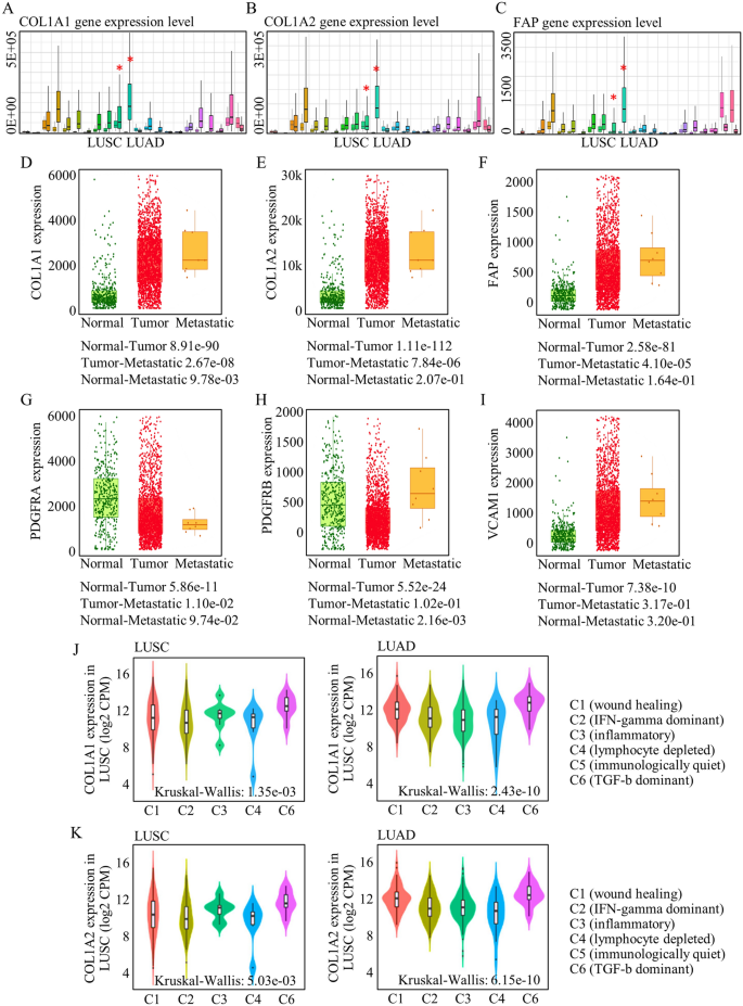
Prior investigations have but to discover the predictive potential of CAF markers for discerning malignancies and metastatic circumstances. This hole in information has been exacerbated by the shortage of environment friendly cell isolation strategies and a scarcity of extremely particular markers. Leveraging our systematic evaluation of assorted markers, now we have recognized COL1A1, COL1A2, FAP, PDGFRA, and PDGFRB as candidates for his or her roles in distinguishing malignancies from regular tissues. The markers of COL1A1 (Fig. 3A), COL1A2 (Fig. 3B), and FAP (Fig. 3C) are considerably extremely expressed in lung most cancers, in comparison with regular tissues, whereas PDGFRA, PDGFRB, and ACTA2 both didn’t be differentially expressed in most cancers tissues, or had been expressed in relative decrease ranges (Fig. S7A-C). Extra particularly and importantly, COL1A1 (Fig. 3D), COL1A2 (Fig. 3E), and FAP (Fig. 3F) had been all extremely expressed in metastatic most cancers tissues, presumably indicated a better likelihood of metastasis when abnormally expressed, which haven’t been reported earlier than. Nevertheless, they had been carefully associated to CAF-based TME capabilities referring to carcinogenesis and tumor metastasis.
CAF correlated with tumor group and indicated malignancy standing. The markers of COL1A1 (A), COL1A2 (B), and FAP (C) are considerably extremely expressed in lung most cancers. COL1A1 (D), COL1A2 (E), and FAP (F) indicated a better likelihood of metastasis. (G–I) The CAF markers of PDGFRA, PDGFRB, and VCAM1 didn’t efficiently differentiate tumor group and regular tissues, or didn’t distinguish metastatic websites. (J,Ok) COL1A1 and COL1A2 had been otherwise enriched in numerous cluster, and their enrichment had been a lot stronger in c-6 group that may most prominently characterize CAF operate.
In distinction, different probably recognized CAF markers didn’t exhibit the capability to discriminate between tumors, and their contributions to assessing survival outcomes remained unclear (see Fig. 3G–I). Consequently, we used COL1A1 and COL1A2 as CAF markers for assessing immune-related cells capabilities utilizing the general public dataset of “TISIDB”. Notably, their enrichment was most pronounced within the c-6 group, which signifies the predominant illustration of CAF performance (see Fig. 3J, Ok).
Survival predicating roles of the consultant COL1A1, COL1A2, and FAP
To outline the roles of every subgroup of CAF inhabitants, we utilized scRNA evaluation of two TCGA datasets (Fig. S6A, B), and fibroblasts constituted the bulk portion in every LUAD pattern dataset (Fig. S6C). To delve deeper into our evaluation, we proceeded to look at the sub-clone clusters amongst lung most cancers sufferers. We categorised fibroblasts based mostly on their respective immune-regulator expression patterns on two TCGA datasets (see Fig. S6D, E). It grew to become evident that distinct fibroblast clusters had been related to various development and general survival outcomes. Given the range within the consultant roles of various CAF markers, we subsequently employed single markers to research their diagnostic and predictive capabilities.
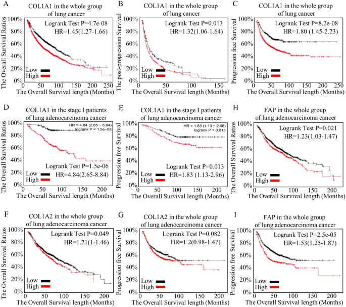
Given {that a} particular CAF marker can discern the malignancy of a tumor and decide the presence or absence of metastasis, we carried out an additional evaluation to look at its predictive capability for survival. Elevated COL1A1 expression was related to shorter general survival (Fig. 4A), post-progression survival (Fig. 4B), and progression-free survival (Fig. 4C) throughout all the spectrum of most cancers sufferers. In subgroup evaluation, COL1A1 proved to be extremely precious in figuring out high-risk teams at an early stage (Fig. 4D, E). Moreover, COL1A2 (Fig. 4F, G) and FAP (Fig. 4H, I) additionally performed vital roles in differentiating survival expectations. Conversely, different elements failed to tell apart variations in survival or variations in disease-free survival. The CAF marker of COL1A1 indicated general survival and disease-free survival extra effectively in early staged lung adenocarcinoma. COL1A2 didn’t distinguish the survival variations of general, development, and post-progression in the entire teams of lung most cancers sufferers (Fig. S7D), Nevertheless, it did really precisely and effectively in early staged lung adenocarcinoma (Fig. S7D). As to FAP, though it may point out survival expectance in general survival, and the progression-free of stage I lung adenocarcinoma, however FAP didn’t outline any distinction in progression-free of different teams of lung adenocarcinoma (Fig. S7E).
Survival predicating roles of the consultant COL1A1, COL1A2, and FAP. As the particular CAF marker may distinguish the malignant state of the tumor and determine the presence or absence of metastasis, we additional analyzed its function in predicting survival. Larger COL1A1 expression pointed to shorter general survival (A), post-progression survival (B), and development free survival (C) in complete most cancers teams. In subgroup evaluation, COL1A1 vastly helped to outlined high-risk teams at early stage (D,E). Additionally, COL1A2 (F,G) and FAP (H,I) indicated a major function in differentiating survival expectances. Different elements failed to tell apart variations in survival or variations in disease-free survival.
CAF ratios correlated with totally different remedies responses
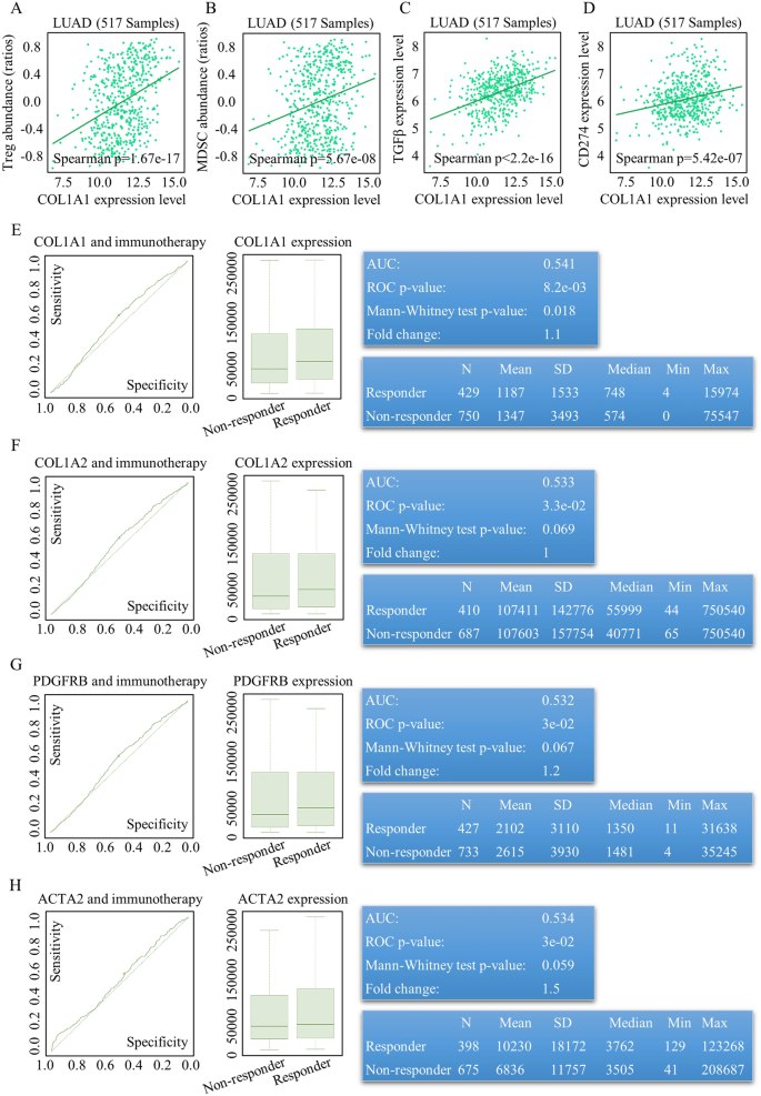
After an intensive evaluation of CAF roles in TME, malignancy defining, survival predication, we lastly assessed the ratios and capabilities of CAF in evaluating the therapy impact. To guage the remedy response referring to totally different CAF markers, we utilized ROC Plotter at https://www.rocplot.com/, and COL1A1 positively correlated with adverse immune regulators, together with inhibitory immune cells (Fig. 5A, B), secretive CAF practical elements (Fig. 5C), and adverse immune regulators (Fig. 5D). In our evaluation, we investigated the predictive worth of CAF markers in relation to immunotherapies throughout varied malignancies. It was noticed that elevated ranges of CAF markers, particularly COL1A1 (Fig. 5E), COL1A2 (Fig. 5F), PDGFRB (Fig. 5G), and ACTA2 (Fig. 5H), had been all related to improved responses to immune remedy. Furthermore, COL1A1 and COL1A2 additionally demonstrated superior predictive talents for the response to TKI (Tyrosine Kinase Inhibitor) therapy, exhibiting sensible ranges of sensitivity and specificity (see Figs. S8 and S9).
CAF ratios variations indicated totally different immune remedy response. COL1A1 positively correlated with inhibitory immune cells [(A,B), T-regulator ratios, raw data could be acquired at http://cis.hku.hk/TISIDB/data_temp/COL1A1_exp_LUAD_TIL_Treg.txt, and MDSC, raw data could be acquired at http://cis.hku.hk/TISIDB/data_temp/COL1A1_exp_LUAD_TIL_MDSC.txt], secretive CAF practical elements [(C), TGFβ expression and secretion, raw data could be acquired at http://cis.hku.hk/TISIDB/data_temp/COL1A1_exp_LUAD_Immunoinhibitor_TGFB1.txt], and adverse immune regulator [(D), CD274 (PD-L1), raw data could be acquired at http://cis.hku.hk/TISIDB/data_temp/COL1A1_exp_LUAD_Immunoinhibitor_CD274.txt]. Elevated CAF markers of COL1A1 (E), COL1A2 (F), PDGFRB (G), ACTA2 (H) indicated higher immune remedy response.
Identification of the relevant scientific and translational values for CAF subgroup
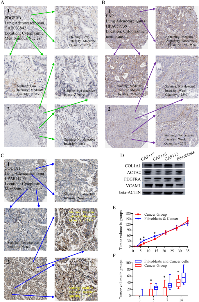
Quite a few scRNA array analyses have make clear the substantial involvement of CAFs within the composition and practical regulation of the TME. To precisely unravel the potential presence and roles of CAFs and their scientific and translational significance, we initiated our investigation by analyzing the immunohistochemistry (IHC) outcomes obtained from the Protein Atlas database. These outcomes had been scrutinized to evaluate the expression patterns of the chosen CAF candidates, together with PDGFRB (Fig. 6A), FAP (Fig. 6B), and COL1A1 (Fig. 6C). These genes usually are not universally expressed in lung most cancers tissues, solely implied for the indicative CAF subgroup, and the full expression degree was not excessive, proving their particular expressing patterns. Additional, we utilized three cell strains of most cancers related fibroblasts, as well as with the management group of fibroblasts, to examine the protein ranges. As to each CAF marker, it might be detected in numerous teams, nonetheless, with totally different expressing depth. PDGFRA and COL1A1 had been universally expressed in three commercially most cancers related fibroblasts. To handle their particular expressing signatures, we detected the relative CAF markers in lung most cancers cell strains, in scientific lung most cancers tissues. COL1A1, ACTA2, and alpha-SMA are comparatively grouped in lung tissues, and PDGFRA, VCAM1 are expressed in each lung tissues and lung most cancers cell strains, exhibiting much less specificity (Fig. S10A). When wanting into the CAF markers extra particularly, COL1A1, ACTA2, and alpha-SMA usually are not considerably totally different in most cancers tissues, in comparison with adjoining lung tissues, partially proving their roles could also be allotted to the TME subgroup, not in complete most cancers tissues or lung tissues. Nude mice implanted with lung most cancers cells alone or related to lung most cancers fibroblasts had been noticed and calculated after injection for 35 days. In vivo examine indicated the upper tumor formation means and extremely proliferative means of tumors triggered by co-embedded CAF teams when performing subcutaneous most cancers cells injection (Fig. 6E, F), however no vital endpoint variations had been seen on the thirty fifth day (Fig. S10B–D).
The relevant scientific and translational utilization for CAF detections and capabilities. The IHC outcomes from protein-atlas had been screened for checking the expressing patterns of enrolled CAF candidates of PDGFRB (A), FAP (B), and COL1A1 (C). These genes usually are not universally expressed in lung most cancers tissues. (D) We utilized three cell strains of most cancers related fibroblasts, as well as with management group of fibroblasts, to examine the protein ranges, and virtually each CAF marker might be detected in numerous teams. (E) In vivo examine indicated the upper tumor formation means and extremely proliferative means of tumors triggered by co-embedded CAF teams when performing subcutaneous most cancers cells injection.


