Sufferers
The multicenter examine was carried out at two hospitals. The examine obtained approval from the Institutional Assessment Board of Zhejiang Most cancers Hospital (Institute 1) and the Institutional Assessment Board of Dongyang Folks’s Hospital (Institute 2), which exempted the requirement for written knowledgeable consent because of the retrospective nature of the examine. Sufferers who met the inclusion standards and have been admitted between June 2019 and January 2022 at Zhejiang Most cancers Hospital have been designated as the invention cohort, whereas these admitted between August 2021 and Might 2023 at Dongyang Folks’s Hospital constituted the validation cohort. Our examine was carried out in accordance with the Declaration of Helsinki.
The examine’s inclusion standards comprised the next: (1) the postoperative histopathological examination confirmed the presence of invasive breast most cancers; (2) clear and full ultrasound photographs of the sufferers’ breast tumors; and (3) availability of complete medical data. Conversely, the exclusion standards included: (1) sufferers who underwent preoperative chemotherapy or radiation remedy; (2) sufferers with breast most cancers offered with a number of lesions; and (3) instances with non-mass lesions that have been troublesome to delineate. The flowchart illustrating affected person choice and dataset development is offered in Fig. 1.
Baseline info was systematically collected for every affected person, comprising key particulars corresponding to age, tumor location, US-reported tumor dimension, Breast Imaging Reporting and Knowledge System (BIRADS) class, US-reported lymph node metastasis, and histopathological options [ER, PR, pathological type, and human epidermal growth factor receptor 2 (HER-2)]. These knowledge have been retrieved from scientific data, postoperative pathology reviews, and immunohistochemistry analyses, guaranteeing a complete analysis and evaluation of the sufferers included on this examine.
Ultrasound imaging acquisition
Ultrasound scans have been carried out utilizing 4 totally different ultrasound devices: the LOGIQ E9 system by GE Healthcare, headquartered in Chicago, Illinois, USA; the Siemens Acuson S2000 system by Siemens Healthineers, situated in Erlangen, Germany; the Toshiba Aplio 500 system manufactured by Canon Medical Methods in Otawara, Japan; and the Philips EPIQ 5 system by Philips Healthcare, headquartered in Amsterdam, Netherlands. These devices have been geared up with probes of varied frequencies, starting from 5 to 18 MHz, to accumulate the biggest long-axis and short-axis ultrasound photographs of the breast lesions. Tumor dimension was decided as the utmost diameter measured within the lengthy axis of the lesion.
In the course of the scanning course of, sufferers have been positioned in a supine or lateral decubitus place, and a water-based gel was utilized to the pores and skin floor to enhance acoustic coupling. The transducer was positioned on the breast area of curiosity, and real-time B-mode imaging was utilized to visualise the lesion. For every affected person, each the long-axis and short-axis planes of the lesion have been scanned and captured to make sure complete protection of the tumor area. The obtained ultrasound photographs have been saved in Digital Imaging and Communications in Medication (DICOM) format to take care of knowledge integrity and facilitate standardized picture evaluation throughout a number of facilities. A centralized image archiving and communication system was utilized to retailer and handle the DICOM photographs securely.
Area of curiosity identification and segmentation
Following ultrasound imaging acquisition, two skilled Sonographers reviewed the saved DICOM photographs to determine the areas of curiosity (ROIs) similar to the breast lesions. The long-axis and short-axis ROIs have been manually delineated utilizing ITK-SNAP software program (Model 3.4.0), respectively, which allowed for exact and constant segmentation of the tumor space. To make sure accuracy and reliability, every ROI segmentation was fastidiously reviewed and validated by a senior Sonographer to reduce inter-observer variability.
Radiomics characteristic extraction and choice
The unique ultrasound photographs have been processed in Python (Model 3.7) utilizing the PyRadiomics package deal. Isotropic pixel factors have been ensured by way of picture preprocessing. Picture normalization (normalize Scale = 25) and resampling (Resample Pixel Spacing = [1, 1, 1]) have been utilized throughout this course of. Then radiomics options have been extracted from the segmented ROIs through the use of pyradiomics package deal, which offered a complete suite of algorithms to quantify numerous image-based options. These options included first-order statistics, texture options, shape-based traits, and wavelet options, all of which contributed to the characterization of the tumor’s spatial heterogeneity and texture patterns. To make sure the robustness of the chosen options, the intra- and inter-observer settlement in ROI segmentation was assessed utilizing the intraclass correlation coefficient (ICC). A excessive ICC worth > 0.75 indicated sturdy settlement, enhancing the reliability and reproducibility of the following radiomics evaluation course of.
Z-score normalization was carried out independently within the discovery and validation cohorts to mitigate the affect of outliers and facilitate the comparability of radiomics options with totally different magnitudes. To determine essentially the most discriminative options for predicting HR standing within the discovery cohort, a rigorous choice course of was employed. Firstly, the intra- and inter-observer settlement amongst Sonographers in ROI segmentation was assessed utilizing the ICC. Subsequently, the Mann–Whitney U take a look at was utilized to check the radiomics options between HR optimistic and HR detrimental subgroups, aiming to determine options with vital variations. Lastly, the least absolute shrinkage and choice operator (LASSO) regression was utilized to additional choose the options that contributed considerably to the predictive mannequin. LASSO regression is a robust method to penalize much less informative options, successfully selling the choice of essentially the most discriminative options for constructing predictive fashions22. By using LASSO regression, the ultimate set of radiomics options was recognized, which performed an important function within the subsequent mannequin development for HR standing prediction.
Mannequin development and validation
With a view to determine threat components related to HR optimistic breast most cancers, each univariate and multivariate regression evaluation have been used. Within the univariate regression evaluation, potential threat components, together with age, tumor location, US-reported tumor dimension, BIRADS class, and US-reported lymph node metastasis have been examined independently. Variables that confirmed a big distinction (p < 0.05) with HR optimistic breast most cancers within the univariate evaluation have been then included within the subsequent mannequin development.
Using the chosen radiomics options, logistic regression fashions have been constructed for each the long-axis and short-axis ultrasound planes, and radiomics rating (Rad-score) was calculated. Moreover, a mixed mannequin utilizing long-axis and short-axis Rad-scores from each planes and scientific threat issue was established. The logistic regression fashions have been skilled utilizing the invention cohort and validated within the validation cohort to evaluate their generalization efficiency. Mannequin efficiency was evaluated based mostly on numerous efficiency metrics, together with sensitivity, specificity, accuracy, and the realm beneath the receiver working attribute (ROC) curve (AUC).
Scientific software
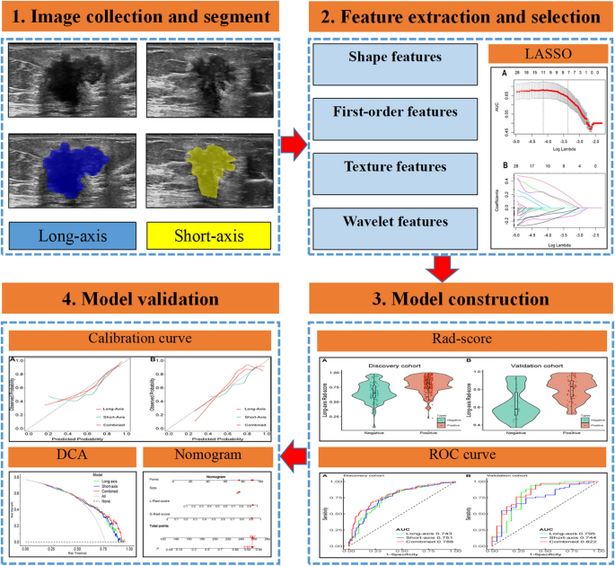
To evaluate its efficiency and reliability, we first evaluated the mannequin’s calibration utilizing a calibration curve. This curve visually compares the expected chances from the mannequin with the precise noticed outcomes. A well-calibrated mannequin would present the factors on the calibration curve near the 45-degree reference line, indicating an excellent settlement between predicted and noticed outcomes. Moreover, we examined the mannequin’s discriminative skill by way of a choice curve evaluation (DCA). The DCA permits us to evaluate the scientific utility of the mannequin by plotting the online profit towards totally different threshold chances. The choice curve supplies insights into the mannequin’s worth in scientific decision-making in comparison with a “treat-all” or “treat-none” technique. The entire mannequin structure is depicted in Fig. 2.
Statistical evaluation
Statistical analyses have been carried out utilizing R software program (Model 4.1.2). For evaluating steady variables between the HR-positive and HR-negative subgroups, the Pupil’s t-test was employed for variables with a standard distribution, whereas the Mann–Whitney U take a look at was utilized for variables with an irregular or unknown distribution. For categorical variables, together with tumor pathological sort, tumor location, and lymph node metastasis, affiliation analyses have been carried out utilizing both the Chi-square take a look at or Fisher’s precise take a look at, relying on pattern dimension and knowledge distribution. A significance degree of two-tailed p < 0.05 was deemed as statistically vital.
Ethics assertion
The analysis involving human members underwent thorough scrutiny and obtained official approval from the Institutional Assessment Board of Dongyang Folks’s Hospital (Approval No. 2024-YX-154) and the Institutional Assessment Board of Zhejiang Most cancers Hospital (Approval No. IRB-2022-548).


