Identification of the immunosenescence-related genes
579 senescence-related genes had been chosen from MSigDB database and 1793 immune-related genes had been obtained from ImmPort. 99 frequent genes amongst them had been acknowledged because the potential ISGs (Supplementary Determine S2A). With STRING on-line database, the PPI evaluation of the potential ISGs was performed and the PPI enrichment p-value was < 1.0e-16 (Supplementary Determine S2B). We additionally employed k-means algorithm on the PPI end result and the 99 ISGs had been usually categorized into two teams. Useful enrichment evaluation indicated that the inexperienced genes had been extra associated to senescence whereas the crimson genes had been extra associated to immune. The PPI community confirmed shut crosstalk between senescence and immune. A lot of the expressions of the 99 ISGs revealed a optimistic correlation in TCGA-STAD (Supplementary Determine S2C). GO organic course of (GO-BP) and KEGG analyses of ISGs proved that these genes had been linked with immune and senescence pathways (Supplementary Determine S2D, E). GSEA confirmed that these organic pathways indicating immunosenescence, together with SASP, mitochondrial biogenesis, glycolysis, and ROS, had been considerably enriched in GC than in regular tissues (Supplementary Determine S2F).
Affirmation of immunosenescence as an indicator of GC on the transcriptomic and genomic degree
To analyze the overall expression situation of immunosenescence in GC, a heatmap of the expression ranges of the ISGs in TCGA-STAD was plotted, displaying that immunosenescence was main considerably enriched in tumor tissues in comparison with adjoining regular ones (Fig. 1A). The ssGSEA was utilized to compute the enrichment degree of the ISGs among the many samples from the TCGA-STAD. Samples whose ssGSEA scores had been greater than the median degree had been thought of because the immunosenescence up-regulation ones and had worse OS (p = 0.025), implying that immunosenescence was considerably linked with the prognosis of GC (Fig. 1B). High 10 variant canonical pathways had been exhibited, indicating that immunosenescence up-regulation was positively linked with adhesion and diapedesis of lymphocytes and granulocytes, and native acute inflammatory response (Supplementary Determine S3A). The standardized scores of eight immune and two stromal cells calculated by MCP-counter technique implied that every one these 10 cells had been extremely expressed in immunosenescence up-regulation group (Supplementary Determine S3B). Though having a better variety of cancer-associated fibroblasts (CAFs) and a better rating of T cell dysfunction, immunosenescence-up sufferers tended to have fewer MDSCs and the M2 subtype of tumor-associated macrophages (TAMs). Additionally they had a decrease T cell exclusion rating and a better cytotoxicity rating (Fig. 1C, D). Moreover, the waterfall plots of mutations revealed that the up-group had increased tumor mutational burden (TMB) (Fig. 1E), and the six genes, specifically TP53, TTN, ARID1A, LRP1B, MUC16, and SYNE1 exhibited the highest 6 highest mutation frequency (Supplementary Determine S3C). Differentially mutated genes had been recognized and HKDC1, MTA1, TNFRSF19 solely mutated in ssGSEA-up set (Supplementary Determine S3D). Primarily based on GISTIC 2.0 software, immunosenescence ssGSEA-down group exhibited comparatively increased G-scores (G = Frequency × Amplitude), implying it had extra SCNAs (Fig. 1F). Heatmaps of the uncooked copy quantity profiles had confirmed this perspective (Supplementary Determine S3E).
Affirmation of senescence as an indicator of GC at transcriptomic and genomic degree. (A) Heatmap of the expression ranges of the ISGs in TCGA-STAD. (B) Survival evaluation of various immunosenescence ssGSEA scores. (C) T cell dysfunction and exclusion standing of various ssGSEA courses. (D) cytotoxicity scores comparability. (E) SNV and (F) SCNA G-score analyses. PPI, Protein–protein interplay; ISGs, immunosenescence-related genes; TCGA-STAD, The Most cancers Genome Atlas abdomen adenocarcinoma; ssGSEA, single-sample gene set enrichment evaluation; SNV, single nucleotide variant; SCNA, somatic copy quantity alteration. ns p ≥ 0.05, *p < 0.05, **p < 0.01, ***p < 0.001, ****p < 0.0001.
Single-cell profiling of immunosenescence inside TME in GC
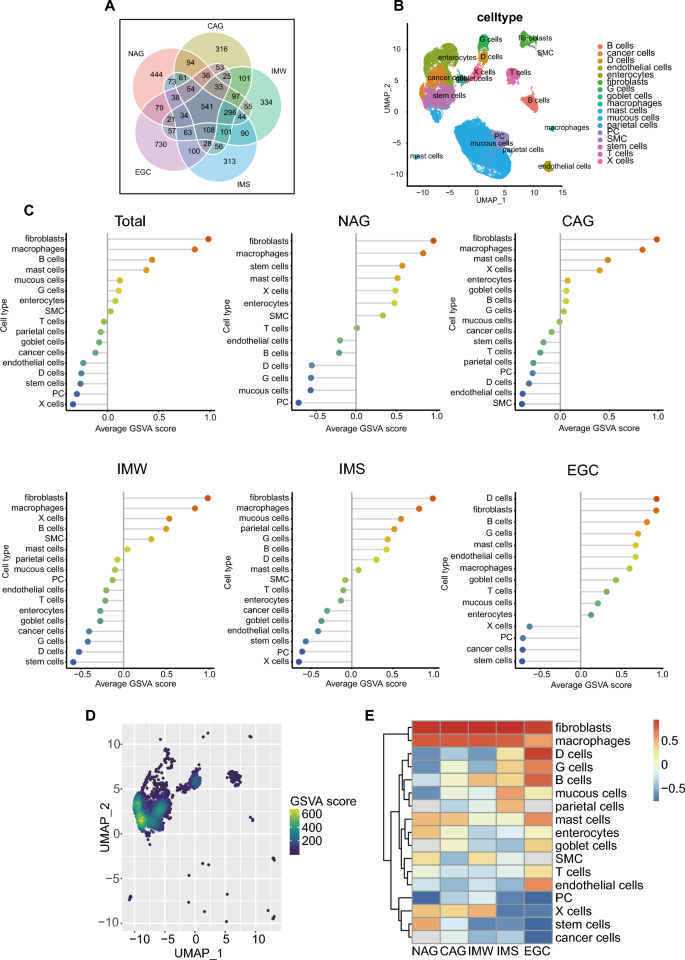
To additional analyze the immunosenescence options of the extent of single-cell, we first researched DRscDB database (Supplementary Determine S4A). ISGs had been enriched in all types of cells corresponding to macrophages and microglia. Contemplating that DRscDB database lacked human abdomen tissues, we downloaded the uncooked knowledge of GSE134520 which contained regular and cancerous gastric tissues to analysis. After important high quality management, 20,685 genes and 48,657 cells (NAG 5639, CAG 19,837, IMW 4165, IMS 15,382, EGC 3634) had been recognized. 2000 HVGs had been recognized as effectively (Supplementary Determine S4B). The Venn diagram of HVGs confirmed that the 5 tissues had 541 frequent HVGs whereas every of them had its personal distinctive HVGs (Fig. 2A). 17 cell sorts had been acknowledged in response to identified marker genes (Supplementary Determine S4C). UMAP of the 17 cell sorts demonstrated that the cells could possibly be categorized into 3 clusters, malignant cells (most cancers cells, stem cells, enterocytes), regular abdomen cells (mucous cells, parietal cells), and different cells, together with immune and stromal cells (Fig. 2B). Immunosenescence GSVA rating was calculated in every type of tissue to painting the dynamic immunosenescence microenvironment in GC growth. Curiously, lollipop diagram, UMAP and heatmap of the GSVA rating confirmed that sure cell sorts consisting of fibroblasts and macrophages, however not the most cancers cells, stem cells, and proliferative cells (PC), had been extremely enriched in immunosenescence signature (Fig. 2C–E). With the event of the illness, D cells, G cells, B cells, T cells, and endothelial cells gained increasingly immunosenescence GSVA scores. The end result prompt that fibroblasts and macrophages, relatively than the most cancers cells or stem cells, primarily contributed to the immunosenescence options in GC.
Single-cell profiling of immunosenescence inside TME in GC. (A) The Venn diagram of HVGs. (B) UMAP of the 17 cell sorts. (C) Common immunosenescence GSVA scores of various cell sorts from complete and 5 tissues. (D) UMAP of immunosenescence GSVA scores in EGC. (E) Heatmap of the dynamic immunosenescence microenvironment in GC growth. TME, tumor microenvironment; HVG, extremely variable gene; UMAP, uniform manifold approximation and projection; GSVA, gene set variation evaluation; EGC, early gastric most cancers; GC, gastric most cancers.
Development of the predictive and prognostic signature
After screening out genes absent in GC immunotherapy cohort (Kim’s cohort), 92 ISGs had been reserved to search out ISGs associated to programmed cell loss of life 1 (PD-1) focused remedy in GC by random forest algorithm. 4 hub genes had been chosen as “ADIPOR2”, “BRAF”, “APOD”, and “C3” (Supplementary Determine S5A). 45 sufferers in Kim’s cohort had been randomly divided into coaching set (n = 30) and validation set (n = 15) to assemble the random forest mannequin primarily based on the hub genes. The mannequin had reached stabilization when the variety of timber (ntree) = 600 (Supplementary Determine S6A). The ROC curve was plotted to research the specificity and sensitivity of the random forest mannequin by “pROC” bundle (Supplementary Determine S6B). The AUC spectacularly attained one, indicating that the 4 hub genes had been considerably linked with the response of GC immunotherapy. Then, the 4 hub genes had been utilized for developing a multivariate Cox regression mannequin to foretell OS of GC sufferers. TCGA-STAD cohort was used because the coaching cohort. To make sure the comparability between totally different datasets, we normalized each cohort and used z-score within the multivariate Cox regression mannequin. The chance rating was calculated as follows: danger rating = 0.303*APOD + 0.064*ADIPOR2-0.038*BRAF-0.011*C3. The mannequin was statistically associated to prognosis (p = 0.02). In line with the median cut-off worth which was equal to -0.004, all of the sufferers had been divided into the high- and low-risk subgroups. To additional analysis the expression of the hub genes at a single-cell degree, TISCH database was used to show the enrichment degree of various cell sorts (Supplementary Determine S6C). APOD was particularly enriched in fibroblasts. ADIPOR2 was barely enriched in myofibroblasts and mast cells whereas BRAF was barely enriched in CD8 + T cells. Final however not least, C3 was enriched in fibroblasts and dendritic cells (DCs).
Exploration of the organic options of the 2 danger teams
We used the limma R software program bundle to acquire DEGs between excessive danger teams and low danger teams. The adjusted p-values of 9196 genes had been < 0.05, amongst which 380 genes had been recognized that absolutely the values of log2 fold change had been > 1. Volcano plot figuring out DEGs amongst two teams was exhibited and APOD was essentially the most vital one (Supplementary Determine S5B). GO and KEGG enrichment analyses indicated that intercellular communication, cell adhesion and development, Calcium signaling pathway, cGMP − PKG signaling pathway, and Apelin signaling pathway had been noticed within the high-risk group of GC (Supplementary Determine S5C, D). Overexpression of apelin and apelin receptor may lead to elevated migration and invasion skills by stimulating migratory protrusion formation and proteolytic exercise by PI3K/AKT and MAPK signaling pathways42. Though cGMP − PKG signaling pathway may work as a protecting signature to intercept proliferation and tumor angiogenesis through inhibiting β-catenin/TCF and SOX9 signaling43, it was the detrimental pathways that performed the dominating position in high-risk group. GSEA evaluation of “hallmark gene units” demonstrated that adipogenesis, epithelial-mesenchymal transition, hypoxia, and KRAS signaling pathway had been enriched within the high-risk group (Supplementary Determine S5E). These signaling pathways play an necessary position in gastric most cancers.
Validation of danger mannequin’s prognostic worth
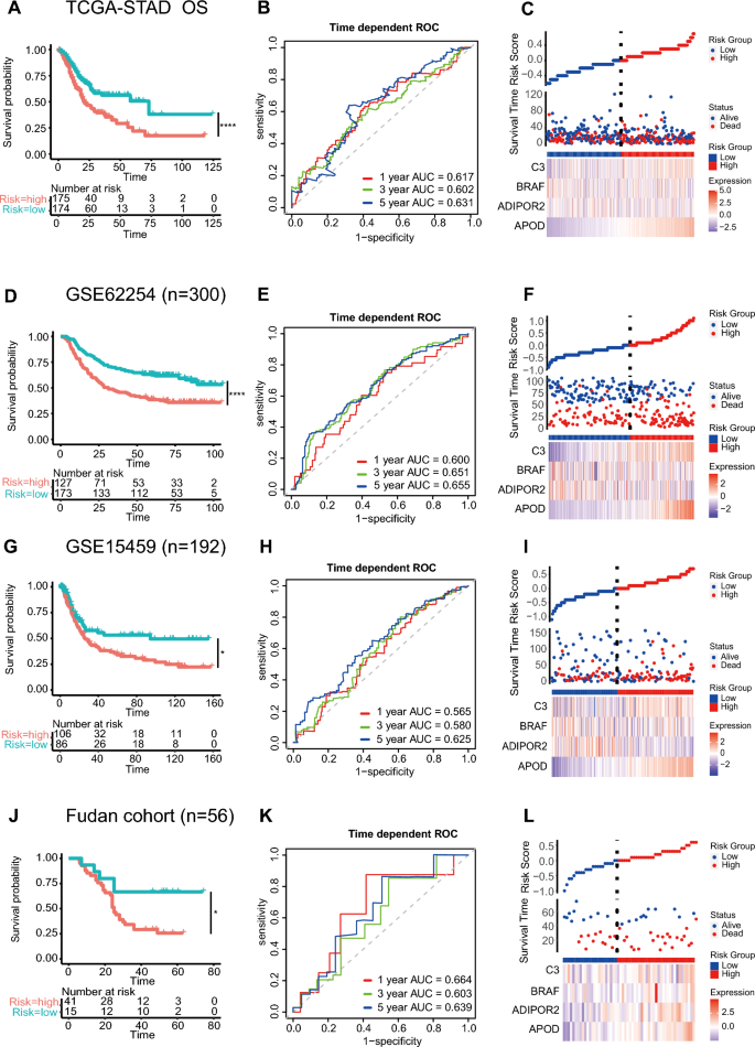
The survival evaluation of TCGA-STAD revealed that the excessive immunosenescence danger rating was related to the poorer OS (p = 0.00022, Fig. 3A), which was reverse to the end result primarily based on senescence danger rating assessed by Zhou et al.44. This discovering forged gentle on the significance of immune for senescence and the crosstalk between them. The time-dependent ROC curve evaluation demonstrated a big predicting potential for OS (1-year AUC = 0.617, 3-year AUC = 0.602, 5-year AUC = 0.631, Fig. 3B). With a view to additional discover the affect of danger scores on the prognosis of GC, we drew a scatter plot of hub gene expression and the corresponding survival time in several TCGA-STAD samples (Fig. 3C). To substantiate the robustness of the danger rating in several folks, we additional explored GSE62254 and GSE15459 datasets with full medical info. Comparable outcomes of survival evaluation had been noticed {that a} increased immunosenescence danger rating was associated to a worse OS of GC sufferers (p < 0.0001, p = 0.017, Fig. 3D, G). Within the GSE62254 cohort, the AUCs of 1-, 3-, and 5-year ROC had been 0.600, 0.651, and 0.655 (Fig. 3E), whereas in GSE15459, the 1-, 3-, and 5-year AUCs had been 0.565, 0.580, and 0.625, respectively (Fig. 3H), displaying nice prognostic discrimination of the mannequin primarily based on hub genes. The distribution of the danger rating, the survival standing, and heatmaps exhibiting the expression of the hub genes in GSE62254 and GSE15459 had been introduced as effectively (Fig. 3F, I). Moreover, Fudan cohort with 56 sufferers was explored because the take a look at set. Encouragingly, the prognostic worth of the danger mannequin was validated (p = 0.019, Fig. 3J). The AUCs of 1-, 3-, and 5-year ROC had been all > 0.6 (Fig. 3Okay). Danger rating correlation diagram of Fudan cohort was according to the results of TCGA (Fig. 3L).
Validation of danger mannequin’s prognostic worth. Survival analyses, ROC curves, and danger rating correlation diagrams had been performed in coaching set (A-C) TCGA-STAD, validation units (D-F) GSE62254 and (G-I) GSE15459, and take a look at set (J-L) Fudan cohort, respectively. ROC, receiver working attribute; TCGA-STAD, The Most cancers Genome Atlas abdomen adenocarcinoma. ns p ≥ 0.05, *p < 0.05, **p < 0.01, ***p < 0.001, ****p < 0.0001.
What’s extra, increased immunosenescence danger rating was additionally associated to the more severe development free interval (PFI) (p = 0.00049) and illness particular survival (DSS) (p < 0.0001) in TCGA-STAD cohort (Supplementary Determine S7A). The time-dependent ROC curve evaluation demonstrated a big predicting potential for PFI (1-year AUC = 0.649, 3-year AUC = 0.631, 5-year AUC = 0.601) and DSS (1-year AUC = 0.676, 3-year AUC = 0.634, 5-year AUC = 0.670) as effectively (Supplementary Determine S7B). Danger rating correlation diagrams of PFI and DSS had been related (Supplementary Determine S7C). Though excessive danger rating may predict poorer PFI and DSS in TCGA-STAD, illness free survival (DFS) was not associated to the danger rating in GSE62254 and Fudan cohorts (Supplementary Determine S8).
Correlation between the danger rating and clinicopathological traits
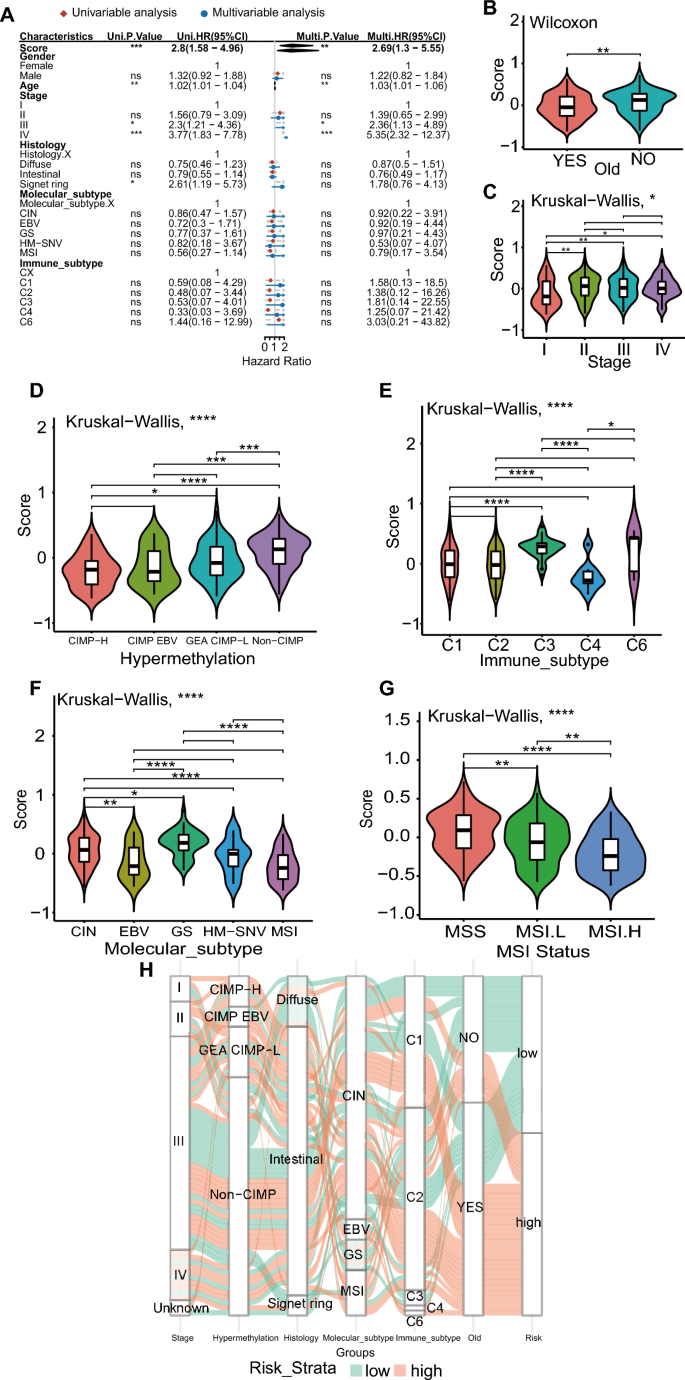
Univariate and multivariate Cox analyzes had been employed to show that the danger rating was an unbiased and strong prognostic issue. A number of clinicopathological options corresponding to gender, age, stage, histology, molecular subtypes, and immune subtypes of TCGA-STAD had been taken under consideration. Amongst them, danger rating, age, stage III and IV had been unbiased prognosis components of GC (P < 0.05, HR > 1, Fig. 4A). Furthermore, vital variations within the danger scores had been noticed amongst clinicopathological subtypes in TCGA cohort. Sufferers older than 60 had been outlined as “outdated”. The chance rating was discovered to be associated to outdated, stage, CpG island methylator phenotype (CIMP)45, immune subtype46, molecular subtype45, and MSI standing (Fig. 4B–G). Curiously, youthful GC sufferers tended to have a better immunosenescence danger rating. Latest research have proved that immunosenescence just isn’t an age-dependent course of, however the dysfunction of the immune system and may embody some traits of low-level continual irritation4. Though some features of immunosenescence could also be associated to getting older, there’s proof that the response to emphasize (whether or not infectious or not) and weight problems are the robust regulators of this course of4. To higher illustrate the traits of the danger rating, an summary of the hyperlinks amongst stage, methylation, histology, molecular subtype, immune subtype, outdated, and particular person affected person danger within the TCGA cohort was represented through the alluvial plot. (Fig. 4H).
Correlation between the danger rating and clinicopathological options. (A) Univariate and multivariate Cox analyses. The relationships between the danger rating and (B) outdated, (C) stage, (D) hypermethylation, (E) immune subtypes, (F) molecular subtypes, and (G) MSI standing. (H) The alluvial diagram confirmed the proportions of high- and low-risk teams in several medical subgroups. MSI, microsatellite instable. ns p ≥ 0.05, *p < 0.05, **p < 0.01, ***p < 0.001, ****p < 0.0001.
Development and verification of signature-based nomograms for OS and PFI
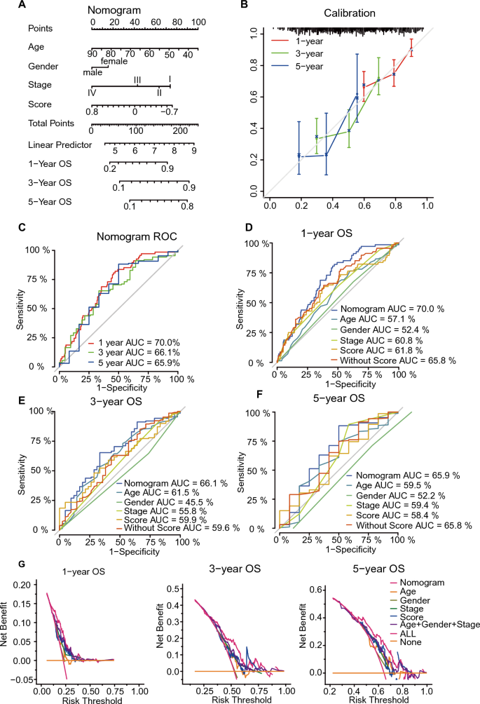
To make this mannequin extra appropriate for medical use, we subsequent constructed a nomogram, which built-in age, intercourse, stage, and danger rating to optimize present metrics for long-term OS prediction by multivariate Cox regression within the TCGA-STAD cohort (Fig. 5A). The nomogram was used to foretell 1-, 3-, and 5-year OS together with medical options and immunosenescence signature. Every affected person was assigned a novel rating primarily based on the constructed nomogram, with increased scores indicating poorer prognosis. The excellence, consistency, and medical utility of the nomogram had been quantified by nomogram calibration curves, time-dependent ROC curves, and DCA.
Development and validation of the signature-based nomogram for OS. (A) Nomogram was developed within the TCGA-STAD cohort primarily based on the danger rating, age, gender, and TNM stage to foretell 1-year, 3-year, and 5-year outcomes. (B) Calibration curves of the nomogram for 1-, 3-, and 5-year OS prediction in TCGA-STAD. (C) Time-dependent ROC curves of the nomogram for 1-, 3-, and 5-year OS prediction in TCGA-STAD. (D-F) Time-dependent ROC curves of the nomogram, age, gender, TNM stage, danger rating, and mannequin with out danger rating for 1-, 3-, and 5-year OS prediction. (G) DCA curves had been carried out to judge the web good thing about the nomogram, age, gender, pathological traits, and danger rating. TCGA-STAD, The Most cancers Genome Atlas abdomen adenocarcinoma; OS, general survival; ROC, receiver working attribute; DCA, determination curve evaluation.
For the nomogram within the coaching cohort, the C-index was 0.673. Nomogram calibration curves demonstrated good settlement between nomogram-predicted OS and precise noticed 1-, 3-, and 5-year OS, additional demonstrating the accuracy of the nomogram (Fig. 5B). The AUCs of 1-, 3- and 5-year ROC had been 0.700, 0.661 and 0.659, respectively (Fig. 5C). Mixed nomograms confirmed the biggest 1-, 3-, and 5-year OS prediction AUCs in comparison with age, intercourse, stage, and fashions with out danger scores, suggesting that incorporating danger scores into the Cox mannequin improved the mannequin’s discriminative energy (Fig. 5D–F). DCA curves confirmed that the nomogram produced a greater web profit in comparison with the person predictors and the mannequin with out danger rating, indicating that the conjunct nomogram may information higher prediction of OS in GC sufferers (Fig. 5G).
As for PFI, we utilized the identical technique to assemble a nomogram within the TCGA-STAD cohort (Supplementary Determine S9A). The C-index of the PFI nomogram mannequin was 0.649. The calibration curves additional demonstrated the accuracy of the PFI nomogram (Supplementary Determine S9B). The AUCs of 1-, 3- and 5-year ROC had been 0.682, 0.647 and 0.657, respectively (Supplementary Determine S9C). The mixed nomograms additionally confirmed the perfect AUCs in comparison with age, intercourse, stage, and fashions with out danger scores in 1-, 3-, and 5-year PFI predictions (Supplementary Determine S9D-F). DCA curves additionally confirmed that the nomogram produced a greater web profit in comparison with different fashions (Supplementary Determine S9G). The same outcomes indicated that incorporating danger scores into the Cox mannequin improved the mannequin’s discriminative energy of each OS and PFI.
Analysis of immune cell infiltration and the capability of predicting immunotherapy response
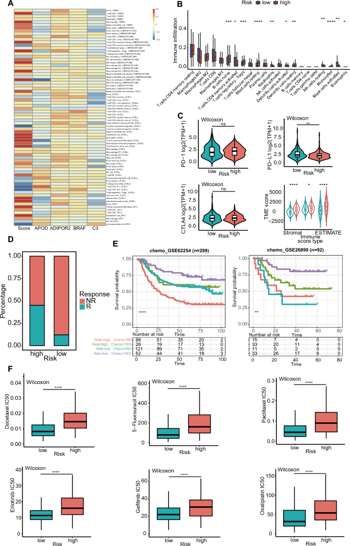
With a view to characterize the immune panorama associated to ISGs, infiltrating immune cell populations had been estimated by 4 algorithms (TIMER, CIBERSORT-ABS, xCELL, and EPIC) (Fig. 6A). Each danger rating and hub genes had been taken under consideration. Correlation evaluation confirmed {that a} increased danger rating indicated elevated abundance of myeloid DCs, endothelial cells, eosinophils, CAFs, granulocyte monocyte progenitors, and hematopoietic stem cells. APOD was positively linked with extra T-helper 1 (Th1) cells and fewer activated mast cells. ADIPOR2 and BRAF had a low optimistic correlation with many of the immune cells. C3 was positively linked with extra CD8 T cells and fewer CD4 T cells and B cells. The CIBERSORT algorithm was employed to judge the correlation between danger rating and 22 immune cells abundance (Fig. 6B). The chance rating was positively associated with the infiltration of naive B cells, regulatory T cells (Tregs), resting mast cells, resting DCs, and monocytes, whereas the opposite efficiency was revealed in relation to danger rating and activated reminiscence CD4 T cells, neutrophils, activated dendritic cells, resting NK cells, activated mast cells. Though high-risk sufferers had comparatively decrease PD-L1 expression, they tended to have increased stromal rating, immune rating, and ESTIMATE rating, indicating a greater immunotherapeutic response (Fig. 6C). Extra responders had been noticed within the high-risk group of Kim’s cohort which acquired anti-PD-1 remedy (Fig. 6D).
The developed signature may predict immunotherapy response and antineoplastic drug sensitivity. (A) Immune infiltration estimations had been carried out utilizing TIMER, CIBERSORT-ABS, xCELL, and EPIC. (B) CIBERSORT was utilized to estimate the fractions of twenty-two tumor-infiltrating immune cells in several danger teams. (C) PD-1, PD-L1, CTLA-4 expression and TME scores of the 2 danger subgroups. (D) The proportion of sufferers with the remedy response to immunotherapy within the high- or low-risk teams. (E) Survival analyses of sufferers from GSE62254 and GSE26899 with totally different danger and chemotherapy statuses. (F) The IC50 values of 6 consultant medication within the two danger teams. IC50, the half-maximal inhibitory focus. TIMER, Tumor Immune Estimation Useful resource; CIBERSORT, cell sort identification by estimating relative subsets of RNA transcripts; ABS, absolute; EPIC, Estimate the Proportion of Immune and Most cancers cells; PD-1, programmed cell loss of life protein 1; PD-L1, programmed cell loss of life ligand 1; CTLA-4, cytotoxic T—lymphocyte antigen 4; ESTIMATE, Estimation of Stromal and Immune cells in Malignant Tumours utilizing Expression knowledge. ns p ≥ 0.05, *p < 0.05, **p < 0.01, ***p < 0.001, ****p < 0.0001.
The developed signature may predict the end result of chemotherapy
In GSE62254 and GSE26899 datasets with info on adjuvant chemotherapy, sufferers had been divided into high-risk and low-risk teams (Fig. 6E). Low-risk sufferers handled with adjuvant chemotherapy had a greatest OS charge. To find out the efficacy of danger scores as a biomarker to foretell the therapy reactions in GC cell traces, we estimated the IC50 values of 198 medication with obtainable from GDSC database. The IC50 values of 122 medication had been discovered associated to the danger classification (Supplementary Desk S3), and most of them had been positively linked with danger scores (Supplementary Determine S10). Consultant medication had been docetaxel, 5-fluorouracil, paclitaxel, erlotinib, gefitinib, and oxaliplatin (Fig. 6F). The end result indicated that high-risk sufferers had been much less delicate to chemotherapy than low-risk ones. Since chemotherapy was nonetheless the first-line remedy for almost all of GC sufferers, the end result was according to the survival analyses.


