Most cancers cells tradition
Immortalized breast most cancers cell vials (MCF-7, SK-BR-3, and MDA-MB-231 cell traces with average to excessive metastatic potential65) had been taken out from storage and cultured in Dulbecco’s Modified Eagle Medium, DMEM, (4.5 g/L glucose, with L-glutamine & phenol pink with out sodium pyruvate, WISENT Inc.) supplemented with 10% v/v of fetal bovine serum (WISENT Inc.) and 1% v/v of streptomycin (WISENT Inc.) in 75 cm2 T-flasks (Thermo Fisher Scientific). Cells had been incubated in an ordinary humidified incubator at 37 °C and 5% CO2. Cells upon reaching a confluency of greater than 70% had been indifferent and passaged. On the day of the experiment (half an hour earlier than the experiments), cells had been indifferent with 0.25% trypsin-EDTA (WISENT Inc.) after three or much less passages. Cells had been quantified for his or her viability with trypan blue with an automatic cell counter (Olympus Life Science). Two to 4 million cells per mL had been grown and used for each experiment.
Design, fabrication, and characterization of microfluidic gadgets
The design of microfluidic gadgets comprises a single constricted channel within the center with sizes corresponding to human microcapillaries to guarantee cell deformation on the constrictions’ entrance (Fig. 10a, b). The microfluidic gadgets had been fabricated utilizing delicate lithography methods. One layer grasp mould was fabricated by spin coating SU-8 photoresist 2015 (Kayaku Superior Supplies) at 1400 rpm for 30 s on a silicon wafer. After spin-coating, the wafer was delicate baked at 65 °C for 1 min adopted by 95 °C for 4 min, and 65 °C for 1 min. Then, the photoresist was uncovered to UV gentle utilizing Karl Suss MA6 Masks Aligner via a beforehand designed and fabricated chromium glass masks (Nanofab, Alberta College) for six s. Subsequent, the post-exposure bake was carried out just like the delicate bake process. Then, the wafer was developed for five min with a SU-8 developer and positioned on a sizzling plate at 150 °C for 20 min to stabilize the SU-8 microstructures. The microscopic photographs of the constricted channels patterned within the grasp mould are proven in Fig. 10c. As well as, the peak of the microchannels was measured to be 28 μm utilizing a floor profilometer (Dektak 8 Stylus Profilometer). After fabricating the silicon grasp mould, polydimethylsiloxane (PDMS) monomer and curing agent had been blended at a ten:1 volumetric ratio. Then, the combination was degasified within the desiccator, poured on the silicon grasp, and thermally cured at 70 °C for two h. The cured PDMS was stripped off from the silicon grasp. Then, the cured PDMS containing the constricted microchannel replicas had been lower to the suitable measurement, punched within the inlets and retailers to acquire the fluidic entry holes, and bounded to a glass slide utilizing oxygen-plasma bonding. The machine fabrication was accomplished by connecting silicon tubing secured with glue to the fluidic entry holes. To characterize the important options of the microfluidic gadgets, shiny subject photographs of the constricted channel of all gadgets captured by an inverted microscope (Nikon Ti Eclipse) had been analyzed. Utilizing a 20× goal, magnified photographs of the constricted channels, constriction entrance, and exit portion had been captured and measured. As Fig. 10a, d present three constricted gadgets every of which has a forty five° tapered entrance on the constricted channel, whose width is corresponding to microcapillary diameters starting from 8 to 12 µm34, that fabricated and used on this research. Furthermore, evaluating to the constricted channels used within the literature16,17,38, measuring cell deformability with cell diameter starting from 13 µm to 26 µm, the fabricated constricted channels within the current research made measuring the single-cell deformability of just about all sizes of the focused most cancers cells possible. In every machine, the width of the channel earlier than the constricted channel is 3 instances bigger than the width of the constricted channel. This assures each the machine fabrication with correct options and maintaining the numerical area sufficiently small for performing the parameters identification. It’s value mentioning that cell samples weren’t filtered previous to infusing into the microfluidic gadgets, however sq. form filters with 50 µm distance from one another had been devised within the inlet of every machine to assist lowering cell mixture on the constricted channel (Fig. 10b).
a Schematic illustration of the microfluidic gadgets design. b Precise picture of the designed one-channel machine and the filters devised within the inlet. c Magnified view of the doorway part of the constricted channels within the fabricated mould utilizing 100× goal. d Magnified view of the constricted channel within the fabricated gadgets utilizing 20× goal. e The fluid velocity magnitude on the mid-plane within the Z route for 20 μL/h movement fee extracted from 3D CFD simulations of every constricted channel
Numerical technique
Lattice Boltzmann technique (LBM)
The entry technique of a single most cancers cell into the constricted channels was modeled utilizing Hemocell open-source code (model 2.4)57. On this code, the fluid is taken into account as an incompressible Newtonian fluid whose movement is described in a Eulerian framework and solved by Lattice Boltzmann Methodology (LBM) applied in Palabos open-source code (model 2.0)66. Extra particularly, a three-dimensional 19-velocity dice lattice scheme (D3Q19) is utilized within the LBM governing equations as follows:
$$start{array}{l}{n}_{i}left(x+{e}_{i}varDelta t,t+varDelta tright)={n}_{i}left(x,tright)-frac{1}{tau }left({n}_{i}left(x,tright)proper. left.-,{n}_{i}^{{eq}}left(x,tright)proper)+{f}_{i}left(x,tright),{rm{for}},i=1,2,ldots ,19end{array}$$
(1)
the place ({n}_{i}left(x,tright)), ({e}_{i}), (varDelta t), (tau) and ({f}_{i}left(x,tright)) are the density distribution operate, the speed vector, time step, leisure time towards the equilibrium distribution ({n}_{i}^{{eq}}), and exterior drive, respectively. At every lattice
web site, the macroscopic fluid density ρ and velocity u could be obtained from the particle density capabilities as follows:
$$start{array}{l}rho left(x,tright)=mathop{sum}limits_{i}{n}_{i}left(x,tright),,{rm{for}},i=1,2,ldots ,19 rho left(x,tright)u=mathop{sum}limits_{i}{n}_{i}left(x,tright){e}_{i},,{rm{for}},i=1,2,ldots ,19end{array}$$
(2)
The numerical domains of the three constricted microfluidic gadgets (proven in Fig. 10a, d) have been created and meshed in a CAD software program (Salome 9.7.0))67 and the fluid passing via every microchannel was modeled utilizing the above described LBM implementation in Hemocell and Palabos. Determine 10e exhibits fluid movement simulation on the mid-plane alongside top route (z-axis) in three microfluidic gadgets. Furthermore, each the channels’ geometry and the geometrical area in x-y aircraft that used for numerical simulation of the cell entry course of within the constricted gadgets had been proven in Fig. 10e.
Cell Membrane Mannequin
The most cancers cell is taken into account as a membrane with a spherical form which is discretized by two-dimensional triangles with springs on the triangles’ edges. The constitutive equations governing the deformation conduct of the most cancers cell embrace a set of forces (the hyperlink drive, the bending drive, the native space drive, and the amount drive) appearing on the cell membrane as described beneath57.
The hyperlink drive acts alongside the sting that connects two adjoining cell membrane vertices is representing the stretch drive on the vertices as outlined beneath:
$${F}_{{rm{hyperlink}}}=-frac{{Ok}_{l}{okay}_{B}T}{P}frac{({L}_{i}-{L}_{0})}{{L}_{0}}left[1+frac{1}{{tau }_{l}^{2}-{(frac{{L}_{i}-{L}_{0}}{{L}_{0}})}^{2}}right]$$
(3)
the place ({Ok}_{l}), ({okay}_{B}), ({rm{T}}), ({L}_{i}), ({L}_{0}) are the hyperlink modulus, the Boltzmann fixed, temperature, the present size of the sting, and the preliminary size of the sting, respectively. (P=7.5,{rm{nm}}) is the persistence-length of the sting, and ({tau }_{l}=3) is the relative growth ratio at which the sting reaches its persistence size57.
The bending drive is outlined when it comes to the change within the angles between the conventional vectors of two adjoining floor components as follows:
$${F}_{{rm{bend}}}=-frac{{Ok}_{b}{okay}_{B}T({theta }_{i}-{theta }_{0})}{{L}_{0}}left[1+frac{1}{{tau }_{b}^{2}-{({theta }_{i}-{theta }_{0})}^{2}}right]$$
(4)
the place ({Ok}_{b}), ({theta }_{i}), and ({theta }_{0}) are the bending modulus, present and preliminary angles between the conventional vectors of the floor components, respectively. ({tau }_{b}) is the limiting angle and is chosen to be (frac{pi }{6}) to forestall unrealistic sharp floor edges57.
The native space drive applies on every floor aspect vertices and represents the response of the aspect to alter of its space as follows:
$${F}_{{rm{space}}}=-frac{{Ok}_{a}{okay}_{B}T}{{L}_{0}}frac{{A}_{i}-{A}_{0}}{{A}_{0}}left[1+frac{1}{{tau }_{a}^{2}-{(frac{{A}_{i}-{A}_{0}}{{A}_{0}})}^{2}}right]$$
(5)
the place ({Ok}_{a}), ({A}_{i}), and ({A}_{0}) are the world modulus, the present and the preliminary space of the triangle, respectively. ({tau }_{a}=0.3) is the world limiting issue to ban floor space adjustments greater than 30%57.
The worldwide quantity drive applies on all vertices of the cell membrane and conserves the amount of the cell.
$${F}_{{rm{quantity}}}=-frac{{Ok}_{v}{okay}_{B}T}{{L}_{0}}left(frac{{V}_{i}-{V}_{0}}{{V}_{0}}proper)left[frac{1}{{tau }_{v}^{2}-{(frac{{V}_{i}-{V}_{0}}{{V}_{0}})}^{2}}right]$$
(6)
the place ({Ok}_{v}), ({V}_{i}), and ({V}_{0}) are the amount modulus the present and the preliminary quantity of the cell membrane, respectively. ({tau }_{v}=0.01) is the amount limiting issue to withstand adjustments within the cell quantity57.
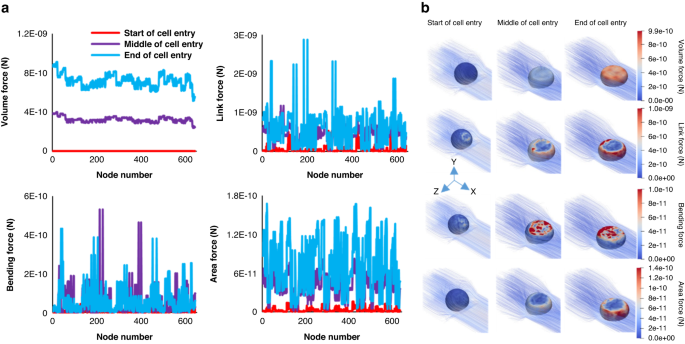
It worths noting that the cell mannequin used on this research consists of 642 nodes on which all of the talked about forces have been utilized at each time step. Determine 11 illustrates the magnitude of the forces at three situations of cell entry course of (begin, center, finish) for 18 µm most cancers cell mannequin coming into the constricted channel of machine #2. This determine exhibits all forces are at their minimal worth earlier than the cell deformation begins. For probably the most or all of the vertices the values of the Quantity forces, the Hyperlink forces, and the Space forces improve because the cell is squeezing to the constriction and attain to their most worth on the finish of the entry course of. For the bending forces most values had been reached through the cell squeezing. Determine 11b depicts the forces act on the cell membrane nodes on the talked about situations by outputting the cell mannequin through the entry course of.
a Magnitude of cell mannequin forces appearing on each node of the cell extracted at three totally different situations (begin, center, finish) throughout cell entry course of for the 18 µm cell coming into the constricted channel of machine #2. b Descriptive photographs of the cell mannequin forces contributing to entrance of the cell into the constriction on the similar situations as half (a)
To realize a practical numerical mannequin on this research, the inner viscosity of the cell was assumed to be totally different from the outside fluid. Due to this fact, a dimensionless parameter named Viscosity Ratio (VR) was thought-about as follows:
$${rm{VR}}=frac{{{rm{Inside}}},{rm{cell}},{rm{fluid}},{rm{viscosity}}}{{{rm{the}}},{rm{fluid}},{{rm{viscosity}}}}$$
(7)
Due to this fact, ({Ok}_{l}), ({Ok}_{b}), ({Ok}_{a}), ({Ok}_{v}), and ({VR}) are dimensionless parameters that must be recognized precisely to allow the numerical mannequin to copy the deformation conduct of the most cancers cell captured within the correspondence experiment.
Fluid-solid interplay (FSI)
Right here, fluid-cell interactions had been modeled with Immerse Boundary Methodology (IBM) which acts as a bridge between Eulerian grids of the fluid and Lagrangian grids of the cell membrane68. Extra particularly, the exerted forces on the cell membrane nodes, decided by the cell’s constitutive equations ((F)), had been unfold on the fluid grids as follows:
$$fleft(x,tright)=int Fleft(q,tright)delta left(x-Xleft(q,tright)proper){dq}$$
(8)
the place (delta) is the Dirac delta operate, (x) is the coordinate of the Eulerian grids, and (X(q,t)) is the place of a cell node with Lagrangian coordinate (q) at time (t).
The rate of the cell membrane nodes (Uleft(Xleft(q,tright)proper)) was obtained from the integral beneath and utilized for updating the positions of the nodes.
$$Uleft(Xleft(q,tright)proper)=int u(x,t)delta left(x-Xleft(q,tright)proper){dx}$$
(9)
the place (uleft(x,tright)) is the speed of the fluid with Eulerian grid (x) at time (t).
Cell–wall interplay
Moreover, repulsive forces had been outlined between the nodes of the cell and microchannel wall to keep away from cell penetration into the microchannel wall to mannequin the conduct of the cell close to the partitions as:
$${vec{F}}_{r}left(dright)={kappa }_{{rep}}frac{{d}_{{lower}}}{d}vec{m},d ,<, {d}_{{lower}}$$
(10)
the place ({kappa }_{{rep}}) is the repulsion fixed, (d) is the gap between the nodes of cell and wall, ({d}_{{lower}}) is the brink of repulsive drive activation, and (vec{m}) is the unit vector pointing from the wall node to the cell node57. The repulsive-force parameters had been fixed in all simulations on this research (Desk 3).
Genetic algorithm (GA)
The talked about parameters which describe the deformation conduct of the most cancers cell had been recognized utilizing the beforehand developed genetic algorithm8. This algorithm advantages from creating the primary inhabitants of measurement 60 utilizing randomly generated multi-digits binary numbers. Each 128 digits of the binary numbers represents one of many parameters and every row of the primary inhabitants represents a set of parameters ({Ok}_{l}), ({Ok}_{b}), ({Ok}_{a}), ({Ok}_{v}), and ({VR}). Afterwards, each two rows of the primary inhabitants had been chosen as dad and mom and crossover has been carried out for the crossover chance increased than a user-defined chance (0.8) for producing youngsters. Then, mutation step was performed for the mutation chance increased than a user-defined chance (0.6) on each digit of the binary numbers that made by crossover. The made youngsters and the mutated ones had been added to the beforehand generated first inhabitants. Then, the binary numbers had been transformed to decimal numbers in line with the higher and decrease bounds of every parameter as offered in Desk 4. Then, utilizing the decimal numbers, numerical simulations of the most cancers cell entry course of had been carried out in all three gadgets benefiting from parallel jobs on supercomputers. It’s value noting that each era consisted of 120 units of parameters, due to this fact, 360 simulations had been carried out concurrently utilizing 2 cores for every and the entry time was saved when the cells absolutely entered the constricted channels. For these simulations, that the entry course of took for much longer than the experimental knowledge, the simulations had been stopped when the entry time reached a user-defined worth (twice of the experimental knowledge). Lastly, the outcomes of the numerical simulations and the experimental knowledge had been in contrast based mostly on the beneath error operate:
$${rm{Error}}=mathop{sum }limits_{n=1}^{{n}_{t}}{E}_{n}=mathop{sum }limits_{n=1}^{{n}_{t}}left|1-{left(frac{{{{rm{ET}}}}^{s}}{{{{rm{ET}}}}^{e}}proper)}_{n}proper|$$
(11)
the place ({E}_{n}),(,{{rm{ET}}}^{s}), and ({{rm{ET}}}^{e}) are the error within the nth machine, numerically calculated entry time, and experimentally measured entry time, respectively. Right here, ({n}_{t}) is the same as 3 for utilizing the entry time at three totally different constricted gadgets.
On the finish, the outcomes for the parameters of the cell constitutive equations had been sorted and the perfect 20 ones added to the preliminary inhabitants of the following era. The algorithm stops if the error stays unchanged after 20 successive generations.
Experimental setup
Earlier than working the experiments, the gadgets had been degassed for as much as sooner or later with Pluronic resolution to keep away from cell adhesion to the channels and washed with a continuing movement of PBS for 20 min. The microfluidic machine was positioned on the stage of a Nikon Ti Eclipse inverted microscope within the experimental setup proven in Fig. 1a. The cell pattern flowing within the media consisting of RPMI-1640 resolution with 20% fetal bovine serum (FBS) (the media density (rho =1020pm 5frac{{{rm{kg}}}}{{{rm{m}}}^{3}}), and the media dynamic viscosity (mu =1.089pm 0.044,{{rm{mPa}}},{rm{s}}))69 with cell viability of %90 or increased was infused into the constricted channel utilizing a syringe pump (Chemyx Inc., USA) at a continuing movement fee of 20 μL/h. Cell samples with the focus of two × 106 cells per mL of media had been ready and used for all experiments. As well as, for the reason that most important focus of the current research is to validate the numerical mannequin of single most cancers cell deformation conduct, solely the info of single most cancers cells that go the constricted channels one by one have been gathered. Extra particularly, the captured knowledge of cell clusters and multiple cells within the constricted channels on the similar time had been put aside from additional evaluation. At this movement fee, the entry time measured for the typical cell measurement is lower than 16 ms. The movement of most cancers cells into the constricted channels has been visualized in shiny subject mode of the inverted microscope utilizing a ×20 magnification goal and recorded utilizing a high-speed digital camera (FASTCAM S1 mannequin, Photon USA, Inc.) at a excessive body fee of 5000 fps with the spatial decision of 512 × 512 pixels. All captured movies and pictures had been analyzed manually utilizing Photron Fastcam Viewer 4 (PFV4) software program to measure cell measurement, entry time, and elongation index which is the ratio of cell size after coming into the constriction to the unique cell size. Determine 1b–d exhibits MDA-MB-231 cell squeezing and coming into the microchannel sizes of 8, 10 and 12 μm, respectively, at 5 situations till completion of most cancers cell entry.


