
On this analysis, all strategies have been carried out in accordance with related tips and rules and all experimental protocols have been accredited by Trita Hospital licensing committee.
Dataset
In step one, the information was collected from Trita Hospital (positioned within the twenty second district of Tehran, Iran). These photos embody T1, T2-tra, T2-sag, T2-cor, DW photos (with 4 sizes of fifty, 500, 1000, and 1500 for B-value) and ADC maps. In response to earlier analysis, T2-tra, T2-sag, DWI (with B-value = 1500), and ADC maps have been chosen for this research as a result of T2 photos present transition zone (TZ) space cancers higher and DW photos present peripheral zone (PZ) space cancers higher. Additionally, rising B-value has a direct relationship with rising accuracy. For that reason, the utmost B-value has been used. (These photos have been produced by an 18-channel gadget with 10 coils receiver). Subsequent, biopsy outcomes have been extracted from medical reviews. The knowledge obtained from the imaging reviews, which included age, antigen degree, dimensions, and quantity of the prostate, was additionally collected. (Since it’s tough to diagnose prostate most cancers at most ranges and this illness has related signs to different illnesses on this space, the scientific opinions of a urology knowledgeable have been used to pick the components. Prostate antigen normally exhibits whether or not the prostate is wholesome or not, however when this issue is blended with the dimensions and dimensions of the prostate, it makes most cancers distinguishable from related illnesses corresponding to prostatitis. Along with this, the age issue can be very influential in most cancers prognosis. For that reason, these components are used concurrently for prognosis and none of them is superior to the opposite.) In response to these reviews, sufferers have been labeled with PI-RADS-V2 scores of 1 to five. Ultimately, 345 sufferers included all this data. (These sufferers visited the imaging middle from October 2020 to December 2021. Knowledgeable consent was obtained from all topics and/or their authorized guardian(s)). The distribution chart of those sufferers is proven in Fig. 1. Additionally, the extracted scientific data contains the:
-
1.
The extent of PSA amongst sufferers was minimal 0.45 and most 230.74 and the typical was 11.96 ng/ml.
-
2.
The prostate quantity amongst sufferers was minimal 12 and most 235 and the typical was 57.21.
-
3.
Sufferers’ age was minimal 39 and most 85 and the typical was 64.05 years.
Pathological knowledge in line with Gleason scoring as two separate numbers are thought-about as enter to introduce the progress degree of the 2 commonest most cancers tissues. In Desk 1, it may be seen that 122 of the sufferers had most cancers in line with the Gleason rating. Additionally, on this desk, the variety of sufferers in line with Gleason grades is introduced.
Desk 2 exhibits the variety of sufferers with most cancers by PIRADS degree. As anticipated, all of the sufferers who had PIRADS 4 and 5 had clinically important most cancers, which signifies the suitable accuracy of imaging strategies in diagnosing clear cancers.
Information preprocessing
Within the pre-processing stage, as a result of solely two sufferers had label 1, this class was eliminated and the variety of sufferers was decreased to 343. Then the size of the photographs turned 150 × 150. Within the final step, the photographs have been normalized. Determine 2 exhibits the sequence of photos of a affected person with stage 4 most cancers. Since neural networks want a considerable amount of knowledge to operate correctly, we had to make use of knowledge augmentation strategies. Medical knowledge may be very priceless and infrequently scarce. As well as, as a result of we wanted picture knowledge and scientific and pathological knowledge on the identical time, the variety of knowledge with these circumstances was very small. The information augmentation technique has been chosen primarily based on earlier works (in these research, it has been proven that the usage of these strategies has elevated the accuracy of the mannequin.) flipping round vertical or horizontal axes11,12,14, rotation11,12,15,16,17, magnification14 and displacement (depth shift)14 has been utilized in research. On this analysis, with the assistance of the ImageDataGenerator operate from the Keras library and the strategies of rotation, symmetry, displacement, and magnification the information have been augmented. Flipping has been achieved in horizontal and vertical instructions. The magnification was 0.2, and the displacement in width and size was 0.2 and 0.2, respectively. Information augmentation has been achieved for all knowledge together with coaching knowledge and take a look at knowledge. As well as, as a result of the variety of knowledge in every class (every illness degree) was not the identical and was far more at degree 3, the mannequin might be extra inclined to study degree 3 and never carry out precisely, subsequently, to unravel the issue, the variety of photos produced within the knowledge increment is weighted. The scientific and pathological knowledge used on this analysis are quantitative and qualitative variables. Quantitative variables have been normalized to optimize coaching. Qualitative variables have been ready for evaluation by the one-hot encoding technique.
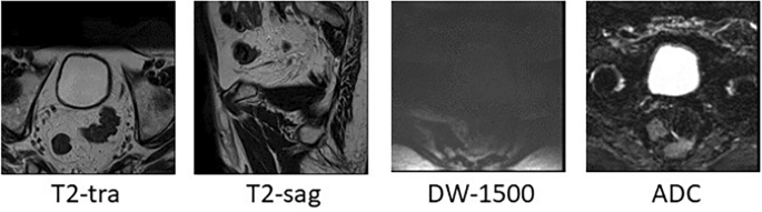
MpMRI of a degree 4 affected person.
Mannequin structure and community coaching
On this analysis, 4 CNN fashions are used concurrently. T2-sag and T2-tra photos, DWI (with B-value = 1500), and ADC maps have been entered into ResNet50. After numerous assessments, these CNNs have been educated with a studying fee of 0.003, batch measurement of 64, in 200 epochs with the Adam optimizer operate. On this analysis, the Relu activation operate has been utilized in all layers of the community besides the final layer which has the duty of classification. Within the final layer of the community, as a result of 4 courses of the issue, the softmax operate is used. Switch studying (ImageNet) has been utilized in every of those networks.
Then the options extracted from every kind of affected person picture (layer earlier than the totally linked layer) have been concatenated collectively and added to the scientific and pathological knowledge. Consequently, a desk was obtained, every document of which was a affected person, during which scientific, pathological data and options extracted from T2-tra, T2sag, DW, and ADC photos have been included. Within the subsequent step, this desk was analyzed by one other neural community. This work made the data obtained from convolutional neural networks to be analyzed with scientific and pathological data. Determine 3 exhibits the general closing mannequin. Determine 4 exhibits the structure of the ANN community.

The used deep studying mannequin.

Remaining neural community structure.