
By
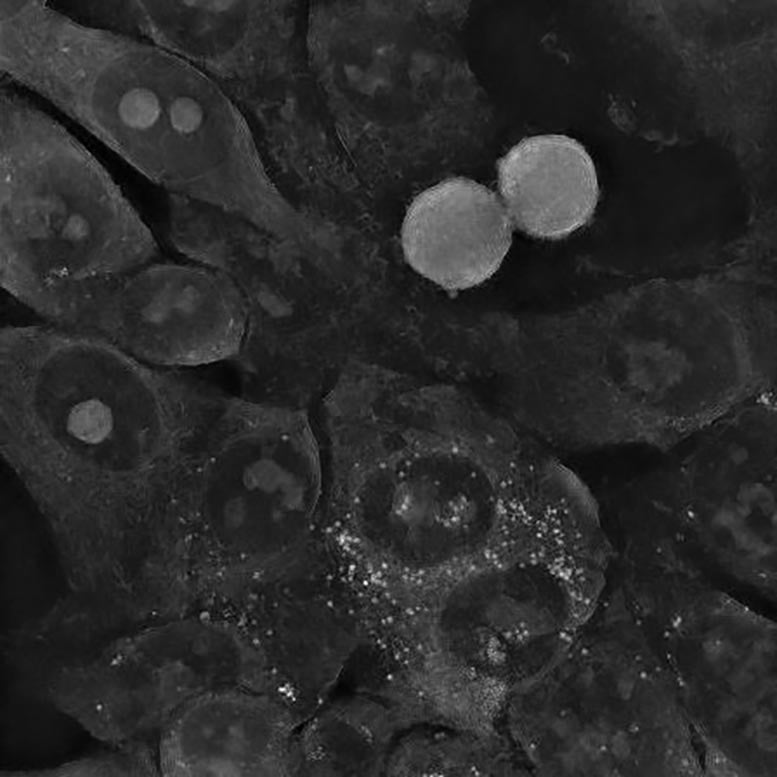
Spherical and star-shaped gold nanoparticles (prime) and colon most cancers cells after approx. 5 hours of publicity to them (backside, respectively). The photograph within the backside left nook proves that, regardless of the small measurement of the spherical nanoparticles, the most cancers cells survived. False colours. Credit score: IFJ PAN
Analysis overturns outdated views on gold nanoparticles, exhibiting bigger, star-shaped ones are simplest in opposition to most cancers, resulting in a mannequin that improves remedy design.
Scientists have beforehand assumed that the smaller the nanoparticles used to struggle the most cancers cells, the sooner they die. The logic behind this concept is that small nanoparticles would merely discover it simpler to penetrate the inside of a most cancers cell, the place their presence would result in metabolic disturbances and finally cell loss of life.
Now, researchers have used a novel microscopic approach to uncover a extra attention-grabbing, advanced image of those interactions.
The examine, not too long ago revealed within the journal Nano Micro Small, was performed by the Institute of Nuclear Physics of the Polish Academy of Sciences (IFJ PAN) and supported by theoretical evaluation carried out on the College of Rzeszow (UR) and theRzeszow College of Know-how.
Colon most cancers cells after interplay with small spherical gold nanoparticles didn’t change their morphology and are nonetheless in a position to divide. Credit score: IFJ PAN
Improvements in Nanoparticle Manufacturing and Testing
“Our institute operates a state-of-the-art medical and accelerator centre for proton radiotherapy. So when stories emerged a couple of years in the past that gold nanoparticles may very well be good radiosensitisers and improve the effectiveness of this kind of remedy, we began to synthesise them ourselves and take a look at their interplay with most cancers cells. We shortly came upon that the toxicity of nanoparticles was not all the time as anticipated,” says Dr. Eng. Joanna Depciuch-Czarny (IFJ PAN), initiator of the analysis and first writer.
Nanoparticles could be produced utilizing a wide range of strategies, yielding particles of various styles and sizes. Shortly after beginning their very own experiments with gold nanoparticles, the IFJ PAN physicists observed that biology doesn’t comply with the favored rule that their toxicity is bigger the smaller they’re. Spherical nanoparticles of 10 nanometres in measurement, produced in Cracow, turned out to be virtually innocent to the glioma cell line studied. Nevertheless, excessive mortality was noticed in cells uncovered to nanoparticles as massive as 200 nanometres, however with a star-shaped construction.
Colon most cancers cells after interplay with small spherical gold nanoparticles didn’t change their morphology and are nonetheless in a position to divide. Credit score: IFJ PAN
Holotomographic Microscopy: A Sport Changer in Most cancers Analysis
Elucidation of the said contradiction turned doable because of using the primary holotomographic microscope in Poland, bought by IFJ PAN with funds from the Polish Ministry of Science and Larger Schooling.
A typical CT scanner scans the human physique utilizing X-rays and reconstructs its spatial inner construction part by part. In biology, the same perform has not too long ago been carried out by the holotomographic microscope. Right here, cells are additionally swept by a beam of radiation, solely not high-energy radiation, however electromagnetic radiation. Its vitality is chosen in order that the photons don’t disturb cell metabolism. The results of the scan is a set of holographic cross-sections containing details about the distribution of refractive index modifications. Since gentle refracts otherwise on the cytoplasm and otherwise on the cell membrane or nucleus, it’s doable to reconstruct a three-dimensional picture of each the cell itself and its inside.
Depciuch-Czarn defined, “In contrast to different high-resolution microscopy strategies, holotomography doesn’t require the preparation of samples or the introduction of any international substances into the cells. The interactions of gold nanoparticles with most cancers cells may due to this fact be noticed straight within the incubator, the place the latter have been cultured, in an undisturbed surroundings, what’s extra with nanometric decision, from all sides concurrently and virtually in actual time.”
Impression of Nanoparticle Form on Most cancers Remedy
The distinctive options of holotomography allowed the physicists to find out the causes of the surprising habits of most cancers cells within the presence of gold nanoparticles. A sequence of experiments was performed on three cell strains: two glioma and one colon. Amongst others, it was noticed that though the small, spherical nanoparticles simply penetrated the most cancers cells, the cells regenerated and even began to divide once more, regardless of the preliminary stress. Within the case of colon most cancers cells, the gold nanoparticles have been shortly pushed out of them. The scenario was totally different for the massive star-shaped nanoparticles. Their sharp suggestions perforated the cell membranes, most probably leading to rising oxidative stress contained in the cells. When these cells may now not deal with repairing the rising harm, the mechanism of apoptosis, or programmed loss of life, was triggered.
Theoretical Modeling and Sensible Purposes
“We used the info from the Cracow experiments to construct a theoretical mannequin of the method of nanoparticle deposition contained in the cells underneath examine. The ultimate result’s a differential equation into which suitably processed parameters could be substituted – in the intervening time solely describing the form and measurement of nanoparticles – to shortly decide how the uptake of the analyzed particles by most cancers cells will proceed over a given time frame,” says Dr. Pawel Jakubczyk, professor on the UR and co-author of the mannequin. “Any scientist can already use our mannequin on the design stage of their very own analysis to immediately slim down the variety of nanoparticle variants requiring experimental verification.”
The flexibility to simply cut back the variety of potential experiments to be carried out means a discount within the prices related to the acquisition of cell strains and reagents, in addition to a marked discount in analysis time (it usually takes round two weeks simply to tradition a commercially obtainable cell line). As well as, the mannequin can be utilized to design better-targeted therapies than earlier than – ones by which the nanoparticles shall be notably properly absorbed by chosen most cancers cells whereas sustaining comparatively low and even zero toxicity to wholesome cells within the affected person’s different organs.
Future Instructions in Nanoparticle Most cancers Analysis
The Cracow-Rzeszow group of scientists is already getting ready to proceed their analysis. New experiments ought to quickly make it doable to increase the mannequin of the interplay of nanoparticles with most cancers cells to incorporate additional parameters, such because the chemical composition of the particles or additional tumor varieties. Later plans additionally embrace supplementing the mannequin with mathematical parts to optimize the efficacy of photo- or proton remedy for indicated mixtures of nanoparticles and tumors.
Reference: “Modeling Absorption Dynamics of Otherwise Formed Gold Glioblastoma and Colon Cells Based mostly on Refractive Index Distribution in Holotomographic Imaging” by Joanna Depciuch, Paweł Jakubczyk, Dorota Jakubczyk, Bartosz Klebowski, Justyna Miszczyk and Magdalena Parlinska-Wojtan, 15 Might 2024, Small.
DOI: 10.1002/smll.202400778