
Introduction
Within the final decade, developments in most cancers therapy have considerably remodeled the oncology panorama with the introduction of precision medication and immunotherapies, demonstrating exceptional success in numerous most cancers sorts comparable to lung cancers and melanoma (1, 2). These developments had been facilitated by early detection and improved characterization of the tumor microenvironment via genomics evaluation and immune profiling, enabling tailor-made therapies. Regardless of these progressions, a number of limitations persist. Remedies for advanced-stage cancers stay restricted, and a few cancers handled within the early levels develop resistance, evading therapies (3). Addressing such challenges stays a formidable job, presumably requiring the invention of latest therapeutic approaches or, extra realistically, a mixture of a number of therapy modalities. Due to this fact, gaining a deeper understanding of how tumor cells and their microenvironment reply to present therapies and assessing the obstacles they encounter can be extremely useful.
With this goal in thoughts, the event and software of superior tumor fashions have develop into indispensable. These fashions permit researchers to work with human cells and function helpful instruments for unraveling the biology of tumors and understanding how therapeutics perform. Over the previous few many years, important strides have been made in enhancing the replication of in vivo tumor environments, particularly via using multicellular 3D fashions (4, 5). These fashions goal to imitate the interactions between numerous cell sorts, together with tumor cells, fibroblasts, and endothelial cells (6). One important good thing about in vitro fashions is the flexibility to recreate tumor environments with various ranges of complexity, allowing a step-by-step method that facilitates a extra complete understanding of the function of various tumor elements and their advanced interplays (7). Nonetheless, one important side that’s usually ignored is the presence of the extracellular matrix mimics (ECM mimics). The ECM mimics can profoundly have an effect on the traits and development of tumors whereas additionally appearing as a barrier that hinders the accessibility of therapeutics. This challenge turns into much more essential when coping with massive molecules or organic brokers comparable to antibodies or oncolytic viruses (OVs). Oncolytic viruses are designed to focus on and destroy tumor cells particularly, with out harming wholesome tissues. The viruses mostly utilized in most cancers virotherapy embrace vaccinia viruses, adenoviruses, herpes simplex viruses, and reoviruses (8). The originality of OVs lies of their multifaceted actions. They exhibit direct cytotoxic exercise by partaking within the lytic virus cycle, ensuing within the amplification of OVs particularly throughout the tumor web site. Subsequent, non-infected tumoral cells may very well be focused with the expression of therapeutic genes by bystander impact, and at last, they may stimulate an immune cell response as overseas microorganisms (9). Now we have developed the oncolytic vaccinia virus TG6002, deleted of thymidine kinase and ribonucleotide reductase to reinforce tumor-selective viral replication (10, 11). TG6002 expresses FCU1, a bifunctional chimeric protein that mixes cytosine deaminase and uracil phosphoribosyltransferase actions and effectively catalyzes the direct conversion of the non-cytotoxic prodrug 5-fluorocytosine (5-FC) into the chemotherapeutic compound 5-fluorouracil (5-FU) (12). TG6002 has lately entered scientific improvement in sufferers with superior gastrointestinal tumors (NCT03724071, NCT04194034). These scientific trials are section 1 research evaluating the security and tolerability of multiple-ascending doses of TG6002 administered intravenously together with oral 5-FC. Though a number of OVs have been evaluated in scientific trials within the final 20 years, solely Talimogene laherparepvec (T-VEC) has been accredited by the US Meals and Drug Administration or the European Medicines Company. T-Vec is used to deal with sufferers with unresectable superior melanoma recurrent after preliminary surgical procedure (13, 14). Consequently, enhancing our data of the mechanisms of motion of OVs would assist overcome potential obstacles and design the following era of oncolytic viruses.
On this examine, our main aim was to design a mannequin to evaluate how the extracellular matrix within the tumor microenvironment impacts the efficacy of oncolytic viruses derived from the vaccinia virus (oVV). Right here, we centered on the cytotoxic exercise of oVV both instantly via its replication or not directly with the expression of an lively payload in a matrix-rich tumoral mannequin. Thus, we selected to create millimeter-scale 3D tumor fashions that incorporate genuine extracellular matrix elements, permitting us to evaluate how this aspect of the tumor microenvironment impacts its efficacy. Among the many numerous strategies employed to create superior fashions (15), we utilized 3D bioprinting know-how, enabling the exact association of cells and the extracellular matrix. We established 3D fashions consisting of colorectal tumor cells (HT29), cancer-associated fibroblasts (CAF), and a bio-inspired proliferative bioink (15). The bioink formulation (comprising gelatin, alginate, and fibrinogen) and its ultimate mechanical properties (Younger’s modulus) could be finely adjusted via the modulation of consolidation reactions (transglutaminase, calcium, and thrombin, respectively) (16). As soon as optimized and characterised, these fashions had been used to judge the habits of various oVVs in a fancy 3D cellularized setting. We evaluated the expression, replication, and cytotoxicity of the oVVs in perform of mannequin stiffness. Taken collectively, our outcomes present that the extracellular matrix impacts oVV efficacy and that each virus replication and expression of a therapeutic payload are crucial to attain antitumoral exercise in a matrix-rich setting.
Materials and strategies
Virus
All oncolytic VACVs had been derived from the Copenhagen pressure and are deleted in thymidine kinase (J2R) and within the massive subunit of ribonucleotide reductase (I4L) genes. oVV-GFP::FCU1 expressing an fusion protein between GFP and FCU1 (ΔI4LΔJ2R/GFP::FCU1 VACV) and oVV-GFP expressing GFP (ΔI4LΔJ2R/GFP VACV) had been constructed and characterised beforehand (10, 17). MVA-GFP::FCU1, the modified vaccinia virus Ankara (MVA) expressing fusion protein between GFP and FCU1, was constructed and characterised as described beforehand (18). MVA-FCU1::GFP and recombinant VACVs had been amplified in main hen embryonic fibroblasts (CEFs) and purified. MVA-FCU1 and oVVs virus inventory had been titrated by plaque assay on CEFs and Vero cells respectively.
Tumor mannequin manufacturing
Tumor fashions had been produced via 3D bioprinting (19). 3D bioprinting bioinks was formulated utilizing bovine gelatin (Merck #G1890), very low viscosity alginate (Alpha Aesar, # A18565) and fibrinogen (Merck, # F8630), every dissolved in a single day in DMEM with out calcium (Gibco™ #21068028) at 37°C. Inventory options of 0.2 g/mL gelatin, 0.04 g/mL alginate and 0.08 g/mL fibrinogen had been ready in Dulbecco’s modified eagle’s medium (DMEM with out calcium (Gibco #21068028) with glutamax-1, Gibco #35050061) supplemented with 10% Fetal Bovine Serum (Thermofisher scientific #A3160802), 100 UI/ml Penicillin-Streptomycin (Thermofisher Scientific, #15140122) and 1 µg/ml amphotericin B (Thermofisher scientific, #15290026).
Then, simply earlier than bioprinting, the bioink was formulated as 0.02 g/mL of fibrinogen, 0.02 g/mL of alginate and 0.05 g/mL of gelatin, and seed with cells. To take action, most cancers related fibrobasts (Neuromics, #CAF05) and colorectal most cancers cells (ATCC, HT29) had been trypsinized and resuspended in 0.08 g/mL fibrinogen at a focus of 1.5 106 cell/ml and 5 106 cell/ml, respectively. To 2mL of this suspended cell answer, 4 mL of alginate inventory answer and a couple of mL of gelatin inventory answer. After homogenization, a ten mL sterile cartridge (Nordson EFD, France) was full of the bioink, incubated quarter-hour at 37°C after which half-hour at room temperature (21°C) to stabilize the bioink rheological properties. The cartridge was then loaded in a 6-axis robotic bioprinter (BioAssemblyBot®, Superior Lifescience Options) and used to print standardized 0.3 cm3 bioprinted tissues (1cm*1cm*2mm). A 410 µm diameter, 6.35 mm lengthy needle (Nordson EFD) was used to bioprint at a set velocity of 10 mm/sec.
As soon as bioprinted, the tumor fashions had been consolidated for 90 minutes at 37°C in an answer containing the next elements: 270 mM of CaCl2 (Sigma, France), 40 or 4 mg/mL of transglutaminase (Ajinomoto, #ActivaWM) and 10 U/mL of thrombin (Sigma, #T4648). As soon as the consolidation accomplished, every bioprinted tissue was rinsed thrice with sterile NaCl 0.9% (Versol, #69600501).
The produced tumor fashions had been then cultured in 12-well plates containing 2 mL of RPMI (ATCC, #30–2001) supplemented with 10% FBS, at 37°C in a 5% CO2 incubator.
Greater than 300 fashions had been produced throughout this examine.
DMA measurements
The viscoelastic habits of the tumor mannequin was characterised by frequency sweep experiments in dynamic mechanical evaluation (DMA) in compression mode. These experiments had been carried out with a rotational rheometer (DHR2, TA Devices, Guyancourt, France) with a DMA mode (torque = 0N) utilizing disk-shaped samples and a parallel plate geometry (8 mm). A preliminary examine was carried out to outline the linear viscoelastic area, which corresponds to the displacement vary the place the fabric properties are assumed to be fixed. This area is decided utilizing oscillatory compression experiments with fixed frequency and ranging displacement. Then, dynamic compression exams had been carried out with a frequency vary of 0.1 to 10Hz (i.e. 0.628 to 62.8 rad/s) at a continuing displacement, which is throughout the linear viscoelastic regime. In these dynamic compression exams, tumor mannequin undergoes a periodical mechanical pressure of very small amplitude and of angular frequency following the Equation 1:
Within the case of the generalized Maxwell mannequin, the storage E′(ω) a part of the advanced modulus is expressed by the Equation 2:
the place is Younger’s modulus of the remoted spring. The relief occasions, , and the dimensionless reference parameters stand for the contribution of every department to the worldwide modulus. The general viscosity could be outlined as Equation 3. The time-constant values had been often distributed between the reciprocals of the very best (62.8 rad/s) and the bottom (0.628 rad/s) angular frequencies of the experimental dynamic modulus. The chosen variety of modes was sufficiently excessive to acquire correct becoming, however not too massive to keep away from inconsistent outcomes (e.g., damaging values of ). Virtually talking, this led to three-time constants (m = 2), often spaced on a logarithmic scale between 5 × 10⁻² s and 5 × 10⁻¹ s.
Identification was achieved by fixing the next minimization drawback described by Equation 3:
the place is the storage modulus obtained from the measured information and is the one computed with viscoelastic parameters. is the variety of measurements acquired in the course of the frequency sweep compression check. The optimization process was carried out through the use of the Microsoft Excel Solver (model 2016) with the Generalized Lowered Gradient (GRG) nonlinear fixing technique.
Macroscopic evaluation
Macroscopic observe up of 3D mannequin development and viability was carried out on a stereo fluorescence microscope (Nikon SMZ18) by transmitted gentle and inexperienced fluorescence following staining of viable cells with Calcein cell-permeant dye at 1 µg/mL (Invitrogen™, #C3100MP).
Histological and immunofluorescence characterization
Histological evaluation was made utilizing paraffin-embedded 3D bioprints after fixation in 4% formaldehyde-16.7mM CaCl2 answer. Paraffin embedded 3D bioprints had been then lower in 4µM sections and mounted on slides. Sections had been deparaffinized, hydrated, after which hematoxylin and eosin (H&E) and Masson-Goldner trichrome (Bio-optical) staining had been carried out for pathomorphological evaluation. Immunohistological analyses for Ki-67, EpCam and Cleaved-caspase 3 had been carried out on Bond RXM (Leica). After deparaffinization and rehydration, epitope retrieval was carried out utilizing Bond epitope retrieval answer 2 (Leica, #AR9640). Endogenous peroxidase actions had been blocked by incubating sections in Hydrogen peroxide block answer (Labvision, #TA-125-H2O2Q), for 10 minutes at room temperature. Sections had been saturated with 10% goat serum for 20 minutes. Main rabbit antibodies rabbit anti-Ki67 (LS-Bio, #LS-B13463), mouse anti-EpCam (Cell Signaling know-how, #2929), and rabbit anti cleaved-caspase 3 (Cell Signaling Expertise, #9661) had been incubated 45 min at room temperature. The mouse anti-Epcam antibody was revealed utilizing Publish Main (rabbit anti mouse secondary antibody Leica, #RE7159), adopted by Novolink anti-Rabbit polymer (Leica, #RE7161), the 2 different antibodies had been revealed utilizing Novolink anti-rabbit polymer alone. Lastly, a Tyramide System Amplification step (TSA-Fluorescein, # SAT701001EA) and a counter staining step utilizing bis-Benzimide Hoechst3328 (Sigma-Aldrich, #B-2883) had been realized.
Hypoxia areas of 3D tumor fashions had been investigated utilizing labeling of pimonidazole hydrochloride (PIMO) which kinds secure covalent adducts with thiol in hypoxia. The staining was carried out utilizing Hydroxyprobe Omni Equipment (Hydroxyprobe, #HP1–100Kit) in accordance with the producer’s protocol. Briefly, fashions had been incubated for 3hrs with a development medium supplemented with 200 µM of PIMO, washed two occasions with PBS and glued in a single day with 4% formaldehyde and Cacl2 (16.7 mM). Subsequent, fashions had been dehydrated, paraffin embedded and lower in 4µm sections. The slides had been handled as described beforehand with the Bond RXM aside from the epitope retrieval which was carried out with Bond epitope retrieval answer 1, and the first antibody which was a rabbit IgG anti-Pimonidazole (1/1000; PAb2627, Hydroxyprobe).
Picture evaluation
ImageJ was used for picture evaluation and cluster quantification. Briefly, full shade microscopy photographs had been remodeled into 8-bit black and white photographs and saved. A pixel depth threshold was then utilized to the remodeled picture and the perform “Analyze Particles” utilized to phase all clusters. Clusters had been then sorted out in accordance with their place within the preliminary picture to plot their distribution throughout the bioprinted tumor mannequin.
An infection
Oncolytic virus an infection was carried out with 3D fashions cultivated inside 6-well plate. The 3D fashions had been contaminated 40 days post-printing when cell clusters had been totally established. The tradition medium was lowered to a quantity of three mL making certain it simply lined the floor of the 3D fashions. Subsequent, an outlined quantity of oVV was rigorously administered drop by drop on the highest of every mannequin. The contaminated 3D tumor fashions had been then incubated beneath managed circumstances at 37°C with 5% CO2. Following 3 hours, the tradition medium was replenished to a ultimate quantity of 5mL and at last the fashions had been maintained at 37°C with 5% calf serum throughout. Tradition medium with 5% calf serum was modified each 2 or 3 days to make sure optimum development circumstances. When fashions had been contaminated with oVV-GFP, an infection of 3D tumor fashions was monitored by following GFP expression from the oVV-GFP with a stereo fluorescence microscope. Fashions had been incubated at 37°C and 5% CO2 in the course of the entirety of the experiment.
Viability evaluation
The viability of the 3D bioprinted tumor fashions was assessed by measuring the metabolic exercise of the cells utilizing resazurin discount utilizing CellTiter-Blue assays (Promega, #G8080) Briefly, on the indicated time, the 3D fashions had been transferred in RPMI supplemented with 10% FBS and 20% CellTiter-Blue. After 9 hours of incubation, 100uL of medium was transferred to 96 wells and the discount of resazurin was decided (560/590 nm excitation/emission filters) with a Tecan Elisa Reader. Sign of contaminated 3D fashions had been in comparison with management fashions handled with 10% RPMI medium to calculate the % of viability.
Enzymatic exercise evaluation
Analysis of cytosine deaminase exercise was quantified by measuring the quantity of 5-FU launched within the tradition media. After 4 days of an infection by oVV-GFP::FCU1 at 10^5 and 10^6 PFUs, 1 mM 5-FC (Toronto Analysis Chemical substances Inc., #F589000) was added to the tradition medium. Each 2/3 days, medium was renewed with 5-FC at 1 mM and 5-FC and 5-FU concentrations within the media had been measured by high-performance liquid chromatography. Twenty microliters of media had been analyzed by high-performance liquid chromatography utilizing a cell section of fifty mM phosphoric acid adjusted to a pH of two.1. Outcomes are expressed as the proportion of 5-FU relative to the entire quantity of 5-FC plus 5-FU.
Outcomes
3D bioprinting permits the era of tumor fashions with an setting wealthy in extracellular matrix
The first goal of this examine is to discover novel therapeutic approaches inside a sensible tumor setting. This entails the creation of human cell-based fashions that incorporate extracellular matrix because it performs an important function in cell metabolism and the effectiveness of anti-tumor therapies.
In vitro fashions of colorectal most cancers (CRC) had been meticulously constructed utilizing 3D bioprinting, using HT29 colorectal tumor cells together with colorectal cancer-associated fibroblasts (CAF) to emulate a consultant CRC setting. These co-cultured cells had been exactly layered inside a proprietary bioink that helps cell development and biocompatibility (15, 20). The stiffness of those preliminary fashions was characterised by a 12 kPa Younger’s modulus (obtained via dynamic mechanical evaluation). The fashions had been cultivated beneath managed circumstances for over 35 days and extensively characterised. Dwell cell development, construction, and group had been visualized via calcein staining, whereas immunohistochemistry was utilized to determine markers for cell proliferation, apoptosis, hypoxia, and most cancers phenotypes.
After 35 days of tradition, viable mobile spheroids, labeled with calcein, had been evident within the CRC mannequin (Determine 1A). Nonetheless, an evaluation of the 3D bioprinted fashions, aided by histochemistry, revealed an uneven distribution of cell clusters throughout the hydrogel. Bigger and extra quite a few cell clusters had been noticed on the periphery, significantly throughout the first 1000 µm close to the sting of the construction (Figures 1B1, C). This distribution of cell clusters is in line with prior findings for related bioprinted buildings and is related to a gradual discount in nutrient and oxygen availability (21). The presence of the EPCAM glycoprotein (Determine 1B2), an adenocarcinoma marker, confirmed that these cell clusters primarily consisted of HT29 cells. Masson’s trichrome inexperienced staining means that the encompassing extracellular matrix, consists of collagen. Based mostly on management experiment with gelatin hydrogel in absence of cell (see Supplementary Determine 1), and on literature it’s hypothesized that this collagen was produced by cancer-associated fibroblasts in the course of the preliminary 35 days of tradition (22, 23). Additional investigations are wanted to verify it.
Determine 1 3D-bioprinted tumor fashions characterization. (A) 35 days after 3D printing, fashions had been stained with calcein AM and had been noticed in vibrant subject (A1), beneath fluorescent gentle to detect reside cells (A2) and at greater magnification (A3). (B) Fashions had been fastened, paraffin embedded and sliced. Consecutive sections had been analyzed by Masson’s trichrome staining (B1), EPCAM (inexperienced) (B2), and immunofluorescent staining towards KI67 (purple) and cleaved caspase 3 (inexperienced) (B3). (C) Cluster dimension distribution alongside 10 mm CRC mannequin histological sections, quantified utilizing imageJ software program and particle evaluation. (D) Hypoxia was monitored by brief incubation with pimonidazole adopted by particular immunofluorescent staining at 10 days (D1) or 35 days (D2) post-printing. Nucleus had been counter-stained with DAPI (Blue).
Proliferation and apoptosis within the cell clusters was additionally assessed utilizing Ki67 and Caspase 3 labeling (Determine 1B3). Curiously, hypoxia, monitored via pimonidazole labeling, didn’t differ considerably based mostly on the placement throughout the mannequin however was related to the time elapsed post-printing and the expansion of the clusters (Determine 1D). Notably, at 35 days post-printing, cell clusters exhibited distinct areas with actively proliferating cells on the outer layer and apoptotic cells on the heart of the cluster (Determine 1B3). The hypoxic area was positioned within the intermediate space the place cells had been actively proliferating.
In abstract, the 3D bioprinted CRC fashions efficiently replicated important traits of tumors, together with heterogeneity in cell proliferation, hypoxia, and the presence of a dense extracellular matrix.
Oncolytic virus infects and maintains long run expression in 3D bioprinted CRC fashions
The 3D CRC fashions had been created to facilitate the examine and improvement of virus-mediated most cancers therapies, with a selected concentrate on understanding the influence of the ECM mimics. Within the preliminary section, we evaluated the affect of ECM mimics on the replication and direct killing of oncolytic vaccinia virus (oVV). To realize this, we employed a first-generation GFP-expressing oVV (oVV-GFP), which is engineered to reinforce tumor-selective viral replication by deleting thymidine kinase and ribonucleotide reductase. Moreover, it expresses the GFP protein as a fluorescent marker to observe virus propagation and expression throughout the advanced bioprinted setting.
Relating to the mechanical properties of the 3D CRC fashions, the preliminary Younger’s modulus, representing the stiffness of the fabric, was measured at 12 kPa. This worth falls a little bit greater than the vary outlined for wholesome colorectal extracellular matrix (1 to five kPa) and is barely under the values for CRC extracellular matrix (25 to 50 kPa) (24). To analyze the influence of stiffness on oVV, two CRC mannequin populations with completely different stiffness ranges had been generated. This was achieved by adjusting the gelatin reticulation throughout the bioink via various transglutaminase concentrations throughout mannequin consolidation (4 or 40 mg/mL), leading to ultimate stiffness values of 12 and 46 kPa. Each fashions had been contaminated with oVV-GFP at 40 days post-printing, and the an infection and virus propagation had been monitored via fluorescent microscopy (Determine 2A).
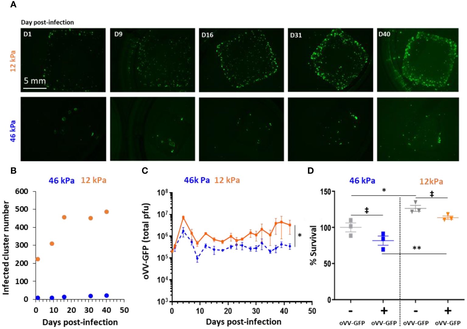
Determine 2 Stiffness of 3D bioprinted tumor mannequin impacts an infection and proliferation of oncolytic Vaccinia Virus. 3D bioprinted tumor fashions with completely different matrix stiffness had been contaminated 40 days post-printing by 10^5 PFU of oVV expressing eGFP. (A) Virus expression was monitored by fluorescent microscopy throughout not less than 40 day post-infection. Matrix with stiffness of 46 kPa (prime) or 12 kPa (backside) had been assessed. (B) Numbers of infected-clusters by oVV-GFP had been quantified utilizing imageJ software program and particle evaluation. (C) Following an infection, supernatant was renewed each 2–3 days and oVV presents within the supernatant was titrated. The orange line and the dashed blue line represented the imply of the entire PFU ± SEM (n=3) for the mannequin with 12 kPa and 46 kPa stiffness respectively. (D) At 42 days post-infection, the viability of the fashions was decided utilizing Celltiter Blue assay. The 100% viability was based mostly on mock-treated fashions. Symbols characterize particular person fashions, and horizontal strains point out the imply ± SEM (n=3). ‡: non-significant ANOVA check (p>0.1). *: important ANOVA check (p<0.02). **: important ANOVA check (p<0.002).
Within the 46 kPa CRC fashions, oVV-GFP efficiently contaminated cell clusters, with GFP clusters noticed as early as day 1 post-infection. Nonetheless, the variety of contaminated clusters at day 1 was minimal (8 clusters), and this quantity didn’t exceed 21 even at day 40 post-infection (Determine 2B). These outcomes counsel that oVV-GFP continued to be expressed at day 40, though with out clear virus diffusion throughout the mannequin.
Within the 12 kPa CRC fashions, GFP clusters had been additionally noticed at day 1 post-infection, and their numbers (224 clusters) had been greater than within the 46 kPa CRC. These numbers continued to extend till day 16, after which within the following days, the variety of contaminated clusters remained secure (round 450). At 40 days post-infection, GFP expression (indicative of virus expression) was extra pronounced and better in comparison with the 46 kPa fashions. In parallel, evaluation by immunostaining of slices derived from 12 kPa fashions after 18 days post-infection confirmed oVV-GFP an infection in solely a portion of the clusters (Supplementary Determine 2).
The quantity of virus launched into the tradition medium of the 2 CRC mannequin populations supported these findings (Determine 2C). Considerably extra virus was launched from the 12 kPa fashions in comparison with the 46 kPa fashions, aligning with the noticed GFP expression throughout the fashions. The kinetics of oVV launch had been related in each fashions, with an preliminary burst of oVV at day 6 post-infection, adopted by a decline in oVV titers. Subsequently, the discharge of oVV reached a plateau, which was maintained for not less than 42 days post-infection when the tradition was concluded. Notably, the plateau of virus manufacturing was decrease within the stiffer 46 kPa fashions, suggesting that virus proliferation and propagation had been hindered by the diploma of extracellular matrix reticulation.
Lastly, cell viability was assessed by measuring their metabolic exercise, with 100% viability set for the 46 kPa non-infected mannequin (Determine 2D). No matter an infection, it was noticed that the cells within the 12 kPa fashions exhibited greater metabolic exercise. Surprisingly, there was no important distinction (ANOVA statistical check) in viability between the contaminated and non-infected fashions, regardless of the mannequin’s stiffness. This means that virus an infection didn’t influence tumor viability, regardless of the continued presence and expression of the virus even 42 days post-infection. Conversely, in a 2D setting, oVV is ready to replicate in HT29 and effectively kills cells with a viability lower than 5% at an MOI of 0.01 5 days submit an infection (Supplementary Determine 3). Doable explanations for the restricted tumor cell killing within the 3D CRC fashions embrace the virus’s restricted capacity to propagate via the extracellular matrix or the resistance of tumor cells in a 3D configuration.
Expression of payload elevated the antitumor effectivity of second era oVV
The primary-generation oncolytic vaccinia virus (oVV) confirmed restricted influence on the viability of HT29 cells throughout the 3D fashions, primarily attributable to challenges in propagating via the bioprinted extracellular matrix. Recognizing the restrictions of direct lysis exercise, a method involving a second-generation oVV expressing a therapeutic gene was explored to reinforce therapeutic effectiveness. On this examine, the examined oVV expresses an enzyme referred to as FCU1 fused with GFP (oVV-GFP::FCU1). FCU1 has the flexibility to transform a non-toxic prodrug, 5-fluorocytosine (5-FC), into the lively chemotherapeutic agent 5-fluorouracil (5-FU), with GFP facilitating direct fluorescence monitoring of virus expression.
Within the current examine, 46 kPa CRC fashions had been contaminated with completely different portions of oVV-GFP::FCU1 (10^6 or 10^5 plaque-forming models) after which cultured for 4 days. Subsequently, the fashions had been handled with 1mM of prodrug 5-FC each 2–3 days (Determine 3A). Whatever the preliminary virus amount used throughout an infection, 5-FC was effectively transformed into 5-FU, with 100% conversion by day 5, showcasing the efficient expression and cytosine exercise of FCU1 (Determine 3B). Nonetheless, after day 5, the enzymatic exercise started to say no, reaching a negligible worth of lower than 5% by day 12 post-infection. This discount in enzymatic exercise corresponded to a lower in virus launch at day 8 post-infection in comparison with fashions contaminated within the absence of 5-FC (Determine 3C). Finally, the an infection by oVV-GFP::FCU1 with the addition of 5-FC induced a big antitumor impact, with a discount in viability to lower than 40% at 18 days post-infection and 14 days submit addition of 5-FC for each virus doses (Determine 3D). As anticipated, neither oVV-GFP::FCU1 nor 5-FC alone had any impact on the viability of the 3D bioprinted fashions even with a better dose of 10^6 PFU. In abstract, these findings reveal that following an infection, oVV-GFP::FCU1 effectively expresses the FCU1 enzyme, resulting in the conversion of 5-FC to 5-FU, ensuing within the loss of life of the 3D bioprinted tumor fashions. This lower in tumor cell content material throughout the fashions subsequently led to a discount in virus proliferation and expression, as reside proliferating cells are required for these processes.
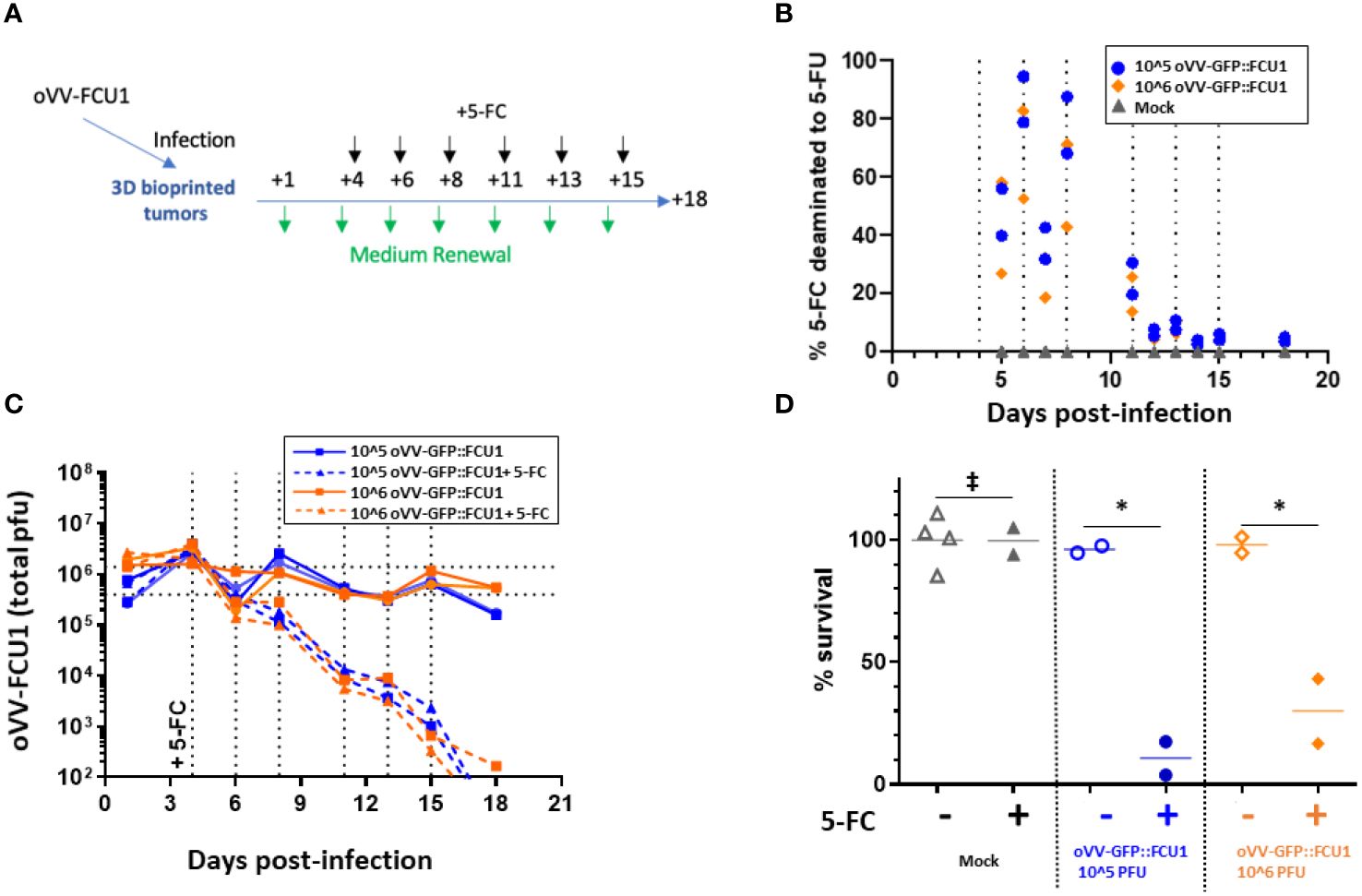
Determine 3 oVV expressing GFP::FCU1 as payload demonstrates environment friendly antitumor cytotoxicity. (A) Schematic depicting the experimental process to judge the antitumor effectivity of oVV-GFP::FCU1. 3D bioprinted-tumor fashions had been contaminated with 10^6 or 10^5 PFU of oVV-GFP::FCU1, the next day enter virus was eliminated and new medium added. Then, 4 days submit an infection 5-FC (1mM) was added in new medium. Each 2/3 days medium with 5-FC was renewed. Experiment was stopped 18 days post-infection and mannequin viability was measured. As management, identical process was utilized with medium with out 5-FC to contaminated and non-infected fashions (for every situation with contaminated fashions (n=2), mock fashions + 5-FC (n=2) and mock fashions (n=4). (B) Focus of 5-FC and 5-FU in mannequin supernatants was decided by high-performance liquid chromatography. The outcomes are offered as % of 5-FU generated from 5-FC with every image representing a person mannequin The % of 5-FC/5-FU conversion was outlined in medium earlier than the medium was renew. (C) Virus within the supernatant was quantified by PFU titration. Every line exhibits the entire PFU measured for particular person fashions. Dotted vertical line indicated the addition of latest medium with 5-FC (D) Viability of the fashions was decided at 18 days post-infection utilizing Celltiter Blue assay. The 100% viability was based mostly on mock-treated fashions. Symbols characterize particular person fashions, and the horizontal line signifies the imply. ‡: non-significant ANOVA check (p>0.9). *: important ANOVA check (p<0.02).
Virus proliferation is important to the antitumor exercise
Given the absence of any antitumor exercise noticed with oVV alone, the examine sought to find out whether or not a replicating virus was important or if the exercise of FCU1 alone is accountable for the antitumoral response. To deal with this, the examine in contrast the antitumoral results of VV-GFP::FCU1 and MVA-GFP::FCU1. MVA-GFP::FCU1 is an attenuated vaccinia virus that can’t replicate in human cells however encodes the GFP::FCU1 gene beneath the same promoter as VV-GFP::FCU1. Each viruses had been utilized to 3D bioprinted tumor fashions at 10^5 PFU, following Determine 3A schedule.
As anticipated, MVA-GFP::FCU1 was incapable of replicating in each circumstances, with or with out 5-FC. Alternatively, the replication of VV-GFP::FCU1 remained fixed with out 5-FC, however, as beforehand noticed (Determine 3C), it decreased after the addition of 5-FC (Determine 4A). Intriguingly, important however average antitumor exercise (85% cell survival) was noticed with MVA-GFP::FCU1 within the presence of 5-FC, whereas extremely environment friendly antitumor exercise (100% cell loss of life) was noticed with the mixture of oVV-GFP::FCU1 and 5-FC (Determine 4B). These outcomes unequivocally reveal that, whereas the replication of oVV-GFP::FCU1 alone is probably not adequate to induce antitumor exercise, it’s certainly crucial. This examine means that in an setting the place optimum oVV proliferation is hindered, comparable to by the extracellular matrix or resistant cells, even minimal virus proliferation can nonetheless play a job by sustaining long-term expression of a therapeutic payload.
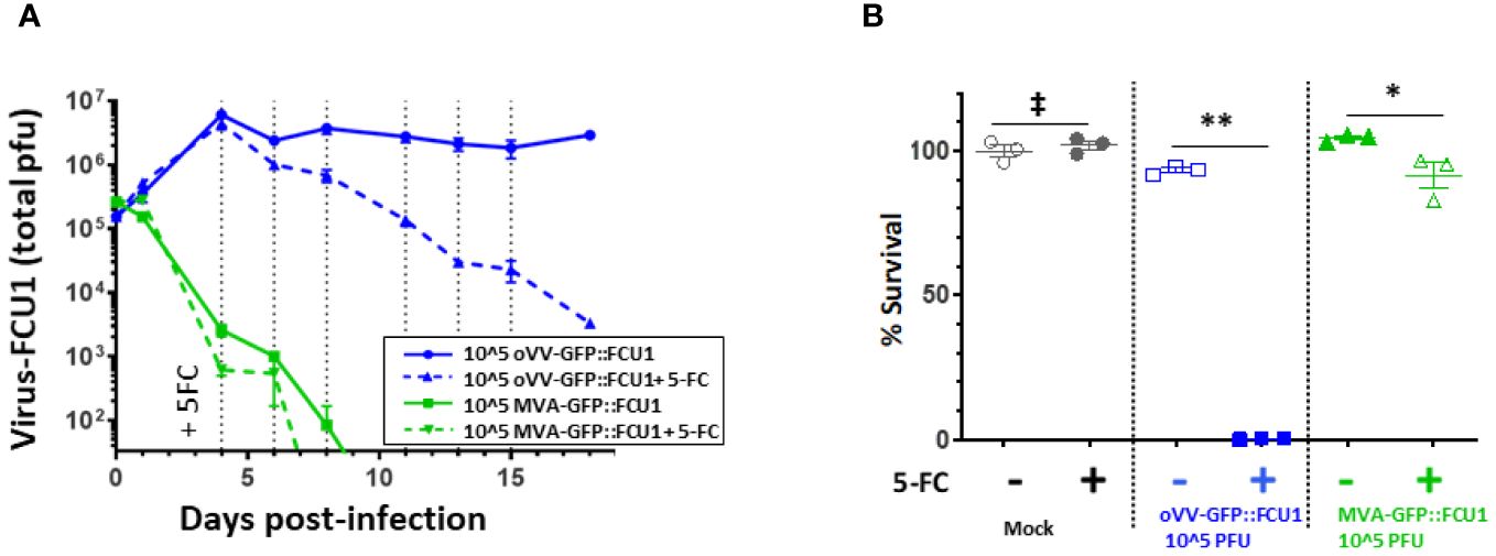
Determine 4 oVV-GFP::FCU1 replication is required for environment friendly antitumoral and bystander impact. (A) 3D bioprinted-tumor fashions had been contaminated by 10^5 PFU of oVV-GFP::FCU1 or MVA-GFP::FCU1 as described in Determine 3A. 5-FC (1mM) was added 4 days submit an infection and each 2/3 days medium was renewed with 5-FC. Virus within the supernatant was quantified by PFU titration. Outcomes are represented because the imply of the entire PFU ± SEM (n=3). As a management, equivalent protocol was carried over with out the addition of 5-FC. Dotted vertical line indicated the addition of latest medium with 5-FC (B) 18 post-infection the viability of the fashions was decided utilizing Celltiter Blue assay. The 100% viability was based mostly on mock-treated fashions. Symbols characterize particular person mannequin, and horizontal line signifies the imply ± SEM (n=3). ‡: non-significant ANOVA check (p>0.9). **: important ANOVA check (p<0.002). *: important ANOVA check (p<0.02).
Dialogue
On this examine, we designed a brand new 3D bioprinted CRC mannequin to analyze the influence of the ECM mimics and its stiffness on oVV effectivity. This seemingly easy query has confirmed difficult to handle with smaller fashions, comparable to multicellular spheroids, the place ECM is proscribed, or in vivo, the place regulating and measuring ECM composition and stiffness are formidable duties.
A number of research have been carried out to develop extra predictive fashions and to maximise the efficacy potential of oncolytic viruses. Preliminary efforts concerned using spheroid tumor fashions, which recapitulate heterogeneous metabolism of tumor cells based mostly on their spatial location throughout the tumor. This contains proliferative cells on the periphery and extra quiescent and necrotic cells towards the core. Research using numerous oncolytic viruses have demonstrated lowered efficacy in such 3D fashions, suggesting potential limitations associated to slower cell metabolism or restricted virus spreading throughout the 3D setting (25, 26). Then, the era of organoid fashions derived from affected person tissues, permits for the exploration of affected person variability and the heterogeneous mobile nature of tumors. Oncolytic viruses derived from Measles and Vaccinia Virus, examined in patient-derived organoids of main breast most cancers, exhibited minimal patient-to-patient variability however effectivity in a three-dimensional configuration was enhanced by payloads expression (27). Conversely, using patient-derived pancreatic tumor organoids to display screen oncolytic adenoviruses highlighted the response variability amongst completely different sufferers (28). These investigations underscore the worth of three-dimensional fashions in gaining understanding of oncolytic virus mechanisms and in creating extra pertinent payloads. Moreover, these research maintain promise for customized therapies, with screening of an oncolytic virus financial institution. Nonetheless, the ECM composition in these fashions is commonly restricted to Collagen or Matrigel, which have considerably decrease stiffness than in vivo tumors (29). Organotypic slices of tumor tissues or tissue explants characterize another method, as they keep the unique elements and group of the tumor. These have been leveraged for evaluating the efficacy and security of oncolytic viruses (30, 31). Nonetheless, sustaining their viability poses a big problem, necessitating environment friendly dealing with and coordination between the working room and the analysis laboratory with a viability usually restricted to at least one week.
Shifting past 3D fashions described above, our examine harnessed the progressive potential of 3D bioprinting to handle the shortcomings in ECM complexity and long-term viability to discover the influence of the ECM mimics and its stiffness on oVV effectiveness. The utilization of 3D bioprinting know-how allowed us to exert management over the fashions’ dimension (reproducible form and dimension), composition (bioink formulation), and, most significantly, their mechanical properties via the fine-tuning of the bioink reticulation. Certainly, the stiffness of ECM in most cancers is commonly greater than in regular tissues and additional will increase because the illness progresses (32). The bioink composition was formulated to imitate as a lot as attainable the standard composition of mammalian tender tissues, i.e. 60–65% water, 16% protein (comprising collagen and different extracellular matrix elements), 1% carbohydrate (33).
Two ranges of stiffness had been applied within the generated fashions: the bottom at 12 kPa, greater than that of wholesome colorectal ECM however decrease than in CRC, and the very best at 46 kPa, falling throughout the vary of CRC Younger’s modulus. Evaluating oVV beneath these two circumstances revealed that stiffness influences oVV development. The influence could be direct, as the upper reticulation of the matrix can bodily impendes oVV. Alternatively, it may be oblique, affecting cell metabolism. We confirmed that the metabolic exercise within the 46 kPa mannequin is decrease than the 12kPa mannequin. On condition that oVV-GFP lacks TK and RR viral enzymes, its replication depends on mobile enzymes. Consequently, decrease cell metabolism within the 46 kPa fashions might restrict oVV replication. It’s noteworthy that, even in 12 kPa mannequin virus, development and antitumoral motion had been restricted. Earlier examine has demonstrated that ECM wealthy in lamin might impaired spreading of oncolytic virus derived from HSV-1 to contaminate cells and in addition its replication after the entry (34). Our examine is to our data the primary to strongly counsel that past the ECM composition, its stiffness impedes the virus propagation.
Additional exploration might contain various the Younger’s modulus to find out if a discount allows enhanced oVV propagation amongst different clusters, or if its propagation limitation is principally attributable to ECM mimics quantity between tumoral clusters. This query might then be addressed with greater cell density or completely different structure that may be simply created via bioprinting. The fashions generated right here might serve to evaluate methods permitting oVV to counteract the ECM mimics, both by expressing degrading enzymes or by mixture with therapies that soften it. Nonetheless, such methods are advanced, as non-selective ECM mimics degradation might result in elevated tumor development or metastasis (35). Therefore, simple screening fashions, because the one described right here, shall be a helpful asset. On one other observe, it will be very tempting to complexify this mannequin to include further sorts of cells, like immune cells, which aren’t solely affected by interplay with tumor cells but in addition with ECM mimics, and to check their habits or their influence on oVV therapies.
The power to take care of the 3D bioprinted mannequin in tradition for over 2 months is of great worth; it permits us to observe oVV an infection for an prolonged interval, which isn’t attainable in classical 2D fashions and even in multicellular spheroids. This led to the stunning statement that virus manufacturing continued for greater than 45 days with out affecting tumoral viability. The dearth of influence on tumor viability may very well be rationalized by the small variety of clusters, and thus cells, which might be reached by oVV attributable to ECM mimics, as defined beforehand (< 5% contaminated cells at d+5 and d+28 post-infection, Supplementary Determine 4). Nonetheless, the continual virus launch and expression are intriguing, contemplating the VV viral cycle in 2D cell tradition is lower than 12 hours and ends in the killing of contaminated cells (36). Consequently, it will be anticipated that after a couple of days, the virus will propagate from cell to cell within the contaminated cluster to induce its destruction after which be stopped because of the absence of residing cells and the impossibility to achieve new cell clusters attributable to ECM mimics. One speculation may very well be that cells are proliferating at the same price than the virus cycle, thus resulting in the alternative of killed cells and sustaining new oVV manufacturing. One other speculation may very well be that in 3D bioprinted fashions, the oVV cycle is modified. It might be longer, not taking simply a number of hours however a number of days. It has been well-documented that tumor cell expression and properties are modified when in 3D cultures in comparison with 2D. For instance, such modifications render them extra immune to therapy (37–39). They could develop into extra immune to cell loss of life induced by oVV and will favor the manufacturing of extracellular enveloped virus. Certainly, two sorts of infectious vaccinia virus particles are sometimes produced in the course of the viral cycle: the intracellular mature virus (IMV) and the extracellular enveloped virus (EEV). The IMV is launched upon cell lysis, whereas EEV launch outcomes from exocytosis or budding via the plasma membrane (40). Characterizing the oVV particles (IMV vs EEV) and evaluating cell expression in a 2D versus 3D bioprinted configuration might present helpful insights to grasp our outcomes.
Lastly, we demonstrated that if a first-generation oVV lacks effectiveness by itself, the induction of a bystander impact via the expression of a therapeutic gene, comparable to FCU1, will destroy the tumor cells. No dose impact was noticed in 5-FC to 5-FU conversion, virus launch, or cell viability. Whereas stunning, this will likely point out {that a} restricted variety of cells could be reached independently to the dose and that payload expression in these cells is adequate. Exploring the efficacy of decrease virus dose mixed with 5-FC shall be attention-grabbing. Nonetheless, utilizing a non-replicating vector derived from the vaccinia virus on the identical dose, we present that expression of FCU1 with the addition of 5-FC was inadequate for eliminating tumor cells. If the bystander impact of FCU1 because of the conversion of produg 5-FC into the potent chemotherapeutic agent 5-FU has been beforehand demonstrated_ENREF_11 (12), the need of virus replication has not been confirmed earlier than. It highlights the added worth of a replicating virus compared to an expression vector, regardless of circumstances wherein replication and propagation of the virus are diminished by a fancy setting. Furthermore, on condition that oVV is able to sustaining a long-term expression of the transgene, that is extremely encouraging for scientific software, particularly in eventualities the place viruses might realistically not attain each tumor cell, and the bystander impact is anticipated to play an important function in therapeutic exercise.
Conclusion and future outlook
On this examine, we efficiently established a 3D tumor mannequin wealthy in ECM mimics, enabling the investigation of obstacles encountered by oncolytic vaccinia viruses (oVVs) throughout their software. We demonstrated that ECM mimics limits their propagation and direct cytotoxic exercise. Nonetheless, the addition of therapeutic payloads like FCU1, together with 5-FC, permits the concentrating on of further tumoral cells and environment friendly antitumor exercise. Importantly, to attain this efficacy, oVV ought to preserve some stage of replication. These outcomes underscore the complementary roles of virus replication and payload expression, emphasizing the important nature of a number of therapeutic modalities for efficient therapy.
Furthermore, this examine serves as a proof of idea, showcasing how the flexibleness of bioprinted 3D fashions will help tackle particular questions concerning the mode of motion of therapies. The additional incorporation of immune cells into this 3D bioprinted mannequin gives an attention-grabbing avenue to sort out questions in regards to the interaction of oVVs, tumor cells, and immunity, with the goal of leveraging immune cell properties to destroy the tumor with out hindering oVVs.
Information availability assertion
The uncooked information supporting the conclusions of this text shall be made accessible by the authors, with out undue reservation.
Ethics assertion
Moral approval was not required for the research on animals in accordance with the native laws and institutional necessities as a result of solely commercially accessible established cell strains had been used.
Creator contributions
CM: Conceptualization, Information curation, Formal evaluation, Funding acquisition, Investigation, Methodology, Challenge administration, Validation, Writing – unique draft, Writing – overview & modifying. EP: Conceptualization, Formal evaluation, Investigation, Methodology, Writing – unique draft, Writing – overview & modifying. AS: Information curation, Writing – overview & modifying. CE: Information curation, Writing – overview & modifying. MN: Investigation, Writing – overview & modifying. BM: Investigation, Writing – overview & modifying. PE: Conceptualization, Information curation, Writing – overview & modifying. J-MB: Conceptualization, Writing – overview & modifying. EQ: Writing – overview & modifying. CZ: Conceptualization, Information curation, Investigation, Writing – unique draft, Writing – overview & modifying.
Funding
The creator(s) declare that no monetary help was obtained for the analysis, authorship, and/or publication of this text.
Battle of curiosity
Creator AS, CE, MN, BM, PE, J-MB, EQ and CZ had been employed by firm Transgene SA.
The remaining authors declare that the analysis was carried out within the absence of any industrial or monetary relationships that may very well be construed as a possible battle of curiosity.
Writer’s observe
All claims expressed on this article are solely these of the authors and don’t essentially characterize these of their affiliated organizations, or these of the writer, the editors and the reviewers. Any product which may be evaluated on this article, or declare which may be made by its producer, will not be assured or endorsed by the writer.
Supplementary materials
The Supplementary Materials for this text could be discovered on-line at: https://www.frontiersin.org/articles/10.3389/fonc.2024.1384499/full#supplementary-material
Supplementary Determine 1 | Masson’s trichrome staining of the bioprinted hydrogel with out cell.
Supplementary Determine 2 | Immunofluorescence evaluation of contaminated 3D-bioprinted tumor fashions. 16 days post-infection fashions had been fastened, paraffin embedded and sliced. Consecutive sections had been analyzed by immunofluorescent staining towards EPCAM (inexperienced) (A), and KI67 (purple) and cleaved caspase 3 (inexperienced) (B) or Vaccinia Virus (Inexperienced) (Anti-VV, Artistic diagnostics # DMAB4487) (C). As management 3D-bioprinted tumor fashions mock-treated had been analyzed utilizing the identical protocol by immunofluorescent staining towards EPCAM (inexperienced) (D), and KI67 (purple) and cleaved caspase 3 (inexperienced) (E) or Vaccinia Virus (inexperienced) (F).
Supplementary Determine 3 | Cytotoxicity of oVV on HT29 monolayer. HT29 tumor cells had been contaminated in suspension by oVV-GFP on the indicated MOI. A complete of three × 10^5 cells/nicely had been plated in 6-well tradition dishes in 2 mL of medium supplemented with 10% FCS. Cells had been then cultured for five days and the viable cells had been counted by trypan blue exclusion utilizing a Vi-Cell Cell Counter (Beckman Coulter). All samples had been analyzed in triplicate. Outcomes are expressed as share of viable cells, 100% akin to mock-infected cells. Values are represented as means ± SD of three particular person determinations.
Supplementary Determine 4 | Quantification of contaminated cells by circulate cytometry. 3D-bioprinted tumor fashions had been contaminated by 1.10^5 pfu of oVV expressing mCherry. An infection was monitored by fluorescent microscopy (A, B), at 5 days post-infection (C) or 28 days post-infection (D) fashions had been dissociated by enzymatic digestion and analyzed by circulate cytometry to quantified mCherry + cells indicating the oVV an infection. P.c of mCherry + cells is indicated within the mCherry+ gate.
References
5. Neufeld L, Yeini E, Pozzi S, Satchi-Fainaro R. 3D bioprinted most cancers fashions: from fundamental biology to drug improvement. Nat Rev Most cancers. (2022) 22:679–92. doi: 10.1038/s41568-022-00514-w
PubMed Summary | CrossRef Full Textual content | Google Scholar
6. Lê H, Deforges J, Hua G, Idoux-Gillet Y, Ponté C, Lindner V, et al. In vitro vascularized immunocompetent patient-derived mannequin to check most cancers therapies. iScience. (2023) 26:108094. doi: 10.1016/j.isci.2023.108094
PubMed Summary | CrossRef Full Textual content | Google Scholar
7. Maulana TI, Kromidas E, Wallstabe L, Cipriano M, Alb M, Zaupa C, et al. Immunocompetent cancer-on-chip fashions to evaluate immuno-oncology remedy. Adv Drug Supply Rev. (2021) 173:281–305. doi: 10.1016/j.addr.2021.03.015
10. Foloppe J, Kempf J, Futin N, Kintz J, Cordier P, Pichon C, et al. The improved tumor specificity of TG6002, an armed oncolytic vaccinia virus deleted in two genes concerned in nucleotide metabolism. Mol Ther oncolytics. (2019) 14:1–14. doi: 10.1016/j.omto.2019.03.005
PubMed Summary | CrossRef Full Textual content | Google Scholar
11. Gallardo F, Schmitt D, Brandely R, Brua C, Silvestre N, Findeli A, et al. Fluorescent tagged vaccinia virus genome permits speedy and environment friendly measurement of oncolytic potential and discovery of oncolytic modulators. Biomedicines. (2020) 8. doi: 10.3390/biomedicines8120543
PubMed Summary | CrossRef Full Textual content | Google Scholar
12. Erbs P, Regulier E, Kintz J, Leroy P, Poitevin Y, Exinger F, et al. In vivo most cancers gene remedy by adenovirus-mediated switch of a bifunctional yeast cytosine deaminase/uracil phosphoribosyltransferase fusion gene. Most cancers Res. (2000) 60:3813–22.
13. Rehman H, Silk AW, Kane MP, Kaufman HL. Into the clinic: Talimogene laherparepvec (T-VEC), a first-in-class intratumoral oncolytic viral remedy. J immunother Most cancers. (2016) 4:53. doi: 10.1186/s40425-016-0158-5
PubMed Summary | CrossRef Full Textual content | Google Scholar
15. Pourchet L, Petiot E, Loubière C, Olmos E, Dos Santos M, Thépot A, et al. Giant 3D bioprinted tissue: Heterogeneous perfusion and vascularization. Bioprinting. (2019) 13:e00039. doi: 10.1016/j.bprint.2018.e00039
16. Marquette CA. Unlocking the potential of bio-inspired bioinks: A collective breakthrough in mammalian tissue bioprinting. Adv Healthc Mater Preprint. (2023). doi: 10.1016/j.bprint.2024.e00351
17. Béguin J, Foloppe J, Maurey C, Laloy E, Hortelano J, Nourtier V, et al. Preclinical analysis of the oncolytic vaccinia virus TG6002 by translational analysis on canine breast most cancers. Mol Ther – Oncolytics. (2020) 19:57–66. doi: 10.1016/j.omto.2020.08.020
PubMed Summary | CrossRef Full Textual content | Google Scholar
18. Erbs P, Findeli A, Kintz J, Cordier P, Hoffmann C, Geist M, et al. Modified vaccinia virus Ankara as a vector for suicide gene remedy. Most cancers Gene Ther. (2008) 15:18–28. doi: 10.1038/sj.cgt.7701098
PubMed Summary | CrossRef Full Textual content | Google Scholar
19. Chastagnier L, el-Kholti N, Essayan L, Thomann C, Courtial EJ, Marquette CA, et al. Deciphering dermal fibroblast habits in 3D bioprinted dermis constructs. Bioprinting. (2023) 32:e00275. doi: 10.1016/j.bprint.2023.e00275
20. Desanlis A, Albouy M, Rousselle P, Thépot A, Santos MD, Auxenfans C, et al. Validation of an implantable bioink utilizing mechanical extraction of human pores and skin cells: First steps to a 3D bioprinting therapy of deep second diploma burn. J Tissue Eng Regener M. (2021) 15:37–48. doi: 10.1002/time period.3148
21. Sbirkov Y, Molander D, Milet C, Bodurov I, Atanasov B, Penkov R, et al. A colorectal most cancers 3D bioprinting workflow as a platform for illness modeling and chemotherapeutic screening. Entrance Bioeng Biotechnol. (2021) 9:755563. doi: 10.3389/fbioe.2021.755563
PubMed Summary | CrossRef Full Textual content | Google Scholar
22. De Martino D, Bravo-Cordero JJ. Collagens in most cancers: structural regulators and guardians of most cancers development. Most cancers Res. (2023) 83:1386–92. doi: 10.1158/0008-5472.CAN-22-2034
PubMed Summary | CrossRef Full Textual content | Google Scholar
24. Nebuloni M, Albarello L, Andolfo A, Magagnotti C, Genovese L, Locatelli I, et al. Perception on colorectal carcinoma infiltration by finding out perilesional extracellular matrix. Sci Rep. (2016) 6:22522. doi: 10.1038/srep22522
PubMed Summary | CrossRef Full Textual content | Google Scholar
25. Tong JG, Valdes YR, Barrett JW, Bell JC, Stojdl D, McFadden G, et al. Proof for differential viral oncolytic efficacy in an in vitro mannequin of epithelial ovarian most cancers metastasis. Mol Ther oncolytics. (2015) 2:15013. doi: 10.1038/mto.2015.13
PubMed Summary | CrossRef Full Textual content | Google Scholar
26. Voros A, Kormos B, Valyi-Nagy T, Valyi-Nagy Ok. Elevated resistance of breast, prostate, and embryonic carcinoma cells towards herpes simplex virus in three-dimensional cultures. ISRN Oncol. (2013) 2013:104913. doi: 10.1155/2013/104913
PubMed Summary | CrossRef Full Textual content | Google Scholar
27. Carter ME, Hartkopf AD, Wagner A, Volmer LL, Brucker SY, Berchtold S, et al. A 3-dimensional organoid mannequin of main breast most cancers to analyze the consequences of oncolytic virotherapy. Entrance Mol Biosci. (2022) 9:826302. doi: 10.3389/fmolb.2022.826302
PubMed Summary | CrossRef Full Textual content | Google Scholar
28. Raimondi G, Mato-Berciano A, Pascual-Sabater S, Rovira-Rigau M, Cuatrecasas M, Fondevila C, et al. Affected person-derived pancreatic tumour organoids determine therapeutic responses to oncolytic adenoviruses. EBioMedicine. (2020) 56:102786. doi: 10.1016/j.ebiom.2020.102786
PubMed Summary | CrossRef Full Textual content | Google Scholar
29. Weigelt B, Ghajar CM, Bissell MJ. The necessity for advanced 3D tradition fashions to unravel novel pathways and determine correct biomarkers in breast most cancers. Adv Drug Supply Rev. (2014) 69-70:42–51. doi: 10.1016/j.addr.2014.01.001
30. Passer BJ, Wu CL, Wu S, Rabkin SD, Martuza RL. Evaluation of genetically engineered oncolytic herpes simplex viruses in human prostate most cancers organotypic cultures. Gene Ther. (2009) 16:1477–82. doi: 10.1038/gt.2009.94
PubMed Summary | CrossRef Full Textual content | Google Scholar
31. Liu Y, Lang F, Xie X, Prabhu S, Xu J, Sampath D, et al. Efficacy of adenovirally expressed soluble TRAIL in human glioma organotypic slice tradition and glioma xenografts. Cell Demise Dis. (2011) 2:e121. doi: 10.1038/cddis.2010.95
PubMed Summary | CrossRef Full Textual content | Google Scholar
33. Lustig JR, Strauss BJG. Encyclopedia of Meals Sciences and Diet. 2nd ed. Caballero B, editor. Oxford: Educational Press (2003) p. 550–7.
34. Valyi-Nagy Ok, Dosa S, Kovacs SK, Bacsa S, Voros A, Shukla D, et al. Identification of virus resistant tumor cell subpopulations in three-dimensional uveal melanoma cultures. Most cancers Gene Ther. (2010) 17:223–34. doi: 10.1038/cgt.2009.73
PubMed Summary | CrossRef Full Textual content | Google Scholar
36. Moss B. Fields Virology. Knipe DM, editor. Philadelphia: Lippincott Williams & Wilkins (2007) p. 2905–46.
37. Costa EC, Moreira AF, de Melo-Diogo D, Gaspar VM, Carvalho MP, Correia IJ. 3D tumor spheroids: an summary on the instruments and strategies used for his or her evaluation. Biotechnol Adv. (2016) 34:1427–41. doi: 10.1016/j.biotechadv.2016.11.002
PubMed Summary | CrossRef Full Textual content | Google Scholar
38. Edmondson R, Adcock AF, Yang L. Affect of matrices on 3D-cultured prostate most cancers cells’ Drug response and expression of drug-action related proteins. PloS One. (2016) 11:e0158116. doi: 10.1371/journal.pone.0158116
PubMed Summary | CrossRef Full Textual content | Google Scholar
39. Abbas ZN, Al-Saffar AZ, Jasim SM, Sulaiman GM. Comparative evaluation between 2D and 3D colorectal most cancers tradition fashions for insights into mobile morphological and transcriptomic variations. Sci Rep. (2023) 13:18380. doi: 10.1038/s41598-023-45144-w
PubMed Summary | CrossRef Full Textual content | Google Scholar